
1. The Human Cell as an Architectural System
A human cell is the basic building block of the body, containing specialized internal structures (organelles) arranged in a highly organized 3‑D layout. This architecture ensures that biochemical reactions occur efficiently and in the correct sequence.
Cells share a universal design:
- Plasma membrane (outer boundary)
- Cytoplasm (internal matrix)
- Organelles (functional compartments)
- Nucleus (genetic control center)
🧱 2. Core Building Blocks of Cellular Architecture
2.1 Plasma Membrane — The Structural Envelope
A dynamic phospholipid bilayer embedded with proteins. Functions:
- Controls entry/exit of substances
- Maintains cell shape
- Hosts receptors and transport channels
- Contains cholesterol for fluidity regulation
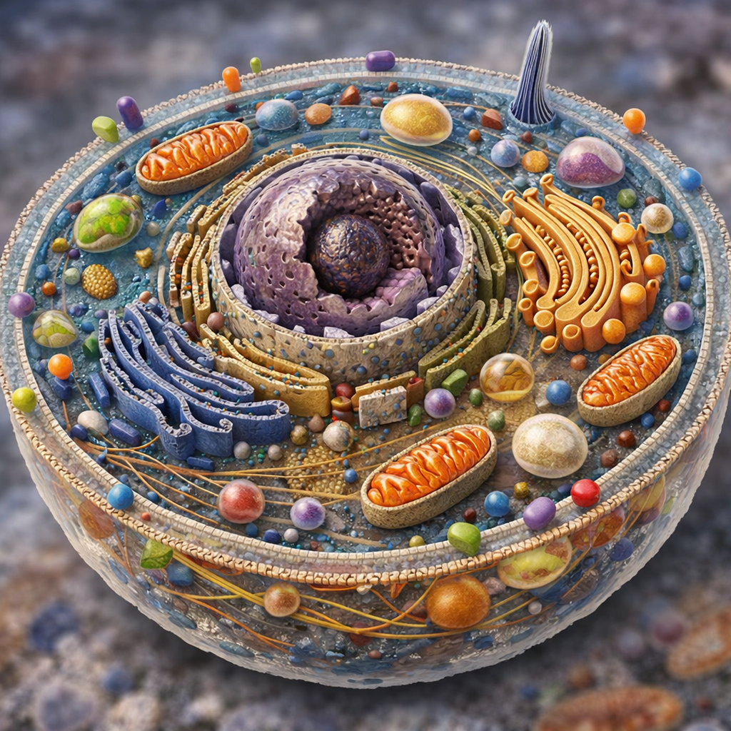
Diagram element: A double-layered boundary with outward-facing hydrophilic heads and inward-facing hydrophobic tails.
2.2 Cytoplasm — The Internal Matrix
A gel-like cytosol containing salts, proteins, sugars, and suspended organelles. Includes:
- Inclusions (glycogen granules, pigments, lipid droplets)
- Cytoskeleton (microtubules, microfilaments, intermediate filaments) for structural support and movement
Diagram element: Semi-fluid background with filament networks.
2.3 Nucleus — The Command Center
Features:
- Nuclear envelope (double membrane with pores)
- Chromatin (DNA + histones)
- Nucleolus (rRNA synthesis)
- Chromosomes (genetic storage units)
Diagram element: Central sphere with a smaller nucleolus inside.
🧩 3. Internal Organelles and Their Diagrammatic Placement
3.1 Ribosomes — Protein Factories
Small molecular machines that translate genetic instructions into proteins. Found:
- Free in cytosol
- Attached to rough ER
Diagram element: Tiny dots on rough ER or floating in cytoplasm.
3.2 Endoplasmic Reticulum (ER) — Manufacturing Network
Two types:
- Rough ER: Ribosome-studded; synthesizes proteins
- Smooth ER: Lipid synthesis, detoxification, Ca²⁺ storage
Diagram element: Folded membrane sheets near nucleus.
3.3 Golgi Apparatus — Packaging & Distribution Hub
Stacks of flattened sacs that modify, sort, and package proteins and lipids for delivery.
Diagram element: Pancake-like stacks with vesicles budding off.
3.4 Mitochondria — Power Plants
Double-membrane organelles generating ATP through cellular respiration. Inner membrane forms cristae for increased surface area.
Diagram element: Oval structures with folded inner membranes.
3.5 Lysosomes & Peroxisomes — Digestive & Detox Units
- Lysosomes: Digest worn-out organelles and pathogens
- Peroxisomes: Break down fatty acids and detoxify chemicals
Diagram element: Small spherical vesicles.
3.6 Vacuoles — Storage Compartments
Store waste and assist in digestion and recycling.
3.7 Primary Cilium — Sensory Antenna
A single hairlike projection used for signal reception.
🧭 4. Diagram Formation: How Internal Structures Are Arranged
4.1 Spatial Logic of Cell Architecture
Electron microscopy reveals that organelles are arranged according to functional flow:
- Nucleus → ER → Golgi → Vesicles → Membrane This ensures efficient protein synthesis, modification, and secretion.
4.2 3‑D Diagrammatic Representation
A typical human cell diagram includes:
- Central nucleus
- Surrounding rough ER
- Smooth ER further outward
- Golgi near ER
- Mitochondria distributed throughout cytoplasm
- Cytoskeleton forming a mesh-like scaffold
- Membrane receptors embedded on the surface
4.3 Ultrastructure Visualization
Electron microscopy shows:
- Ribosomes as dense dots
- ER as layered membranes
- Mitochondria with distinct cristae
- Golgi as stacked cisternae
- Cytoskeleton as fibrous strands
🧪 5. Functional Integration: How Architecture Enables Life
5.1 Protein Flow
- DNA → mRNA (nucleus)
- mRNA → protein (ribosomes)
- Protein → ER → Golgi
- Golgi → vesicles → membrane or secretion
5.2 Energy Flow
Glucose → mitochondria → ATP → drives cellular processes.
5.3 Structural Flow
Cytoskeleton rearranges to:
- Change cell shape
- Enable movement
- Assist in division (mitosis)
🧠 6. Why This Architecture Matters
The cell’s internal design ensures:
- Efficient biochemical reactions
- Controlled communication
- Adaptability
- Survival under stress
- Accurate replication



Be First to Comment