A fully simplified account of structure, energy metabolism, signalling, and disease
Abstract
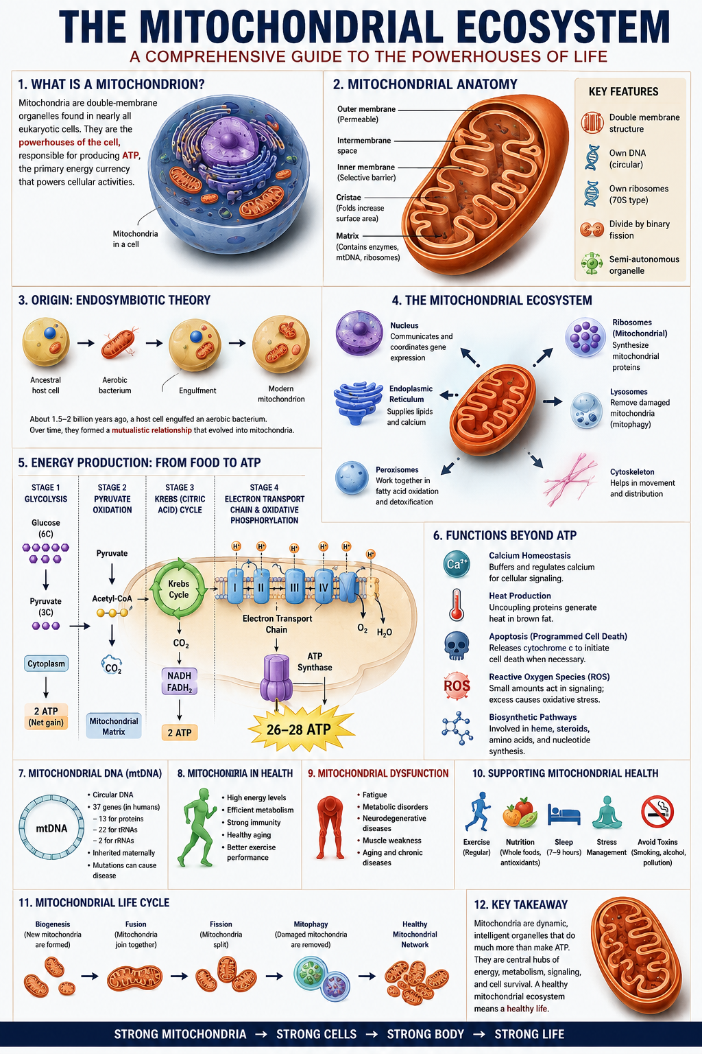
Mitochondria are usually introduced as “the powerhouse of the cell” — true, but radically incomplete. This companion treats the mitochondrion as an ecosystem: a semi-autonomous, genetically distinct, dynamically networked population of structures governing energy conversion, biosynthesis, calcium buffering, redox balance, and the cell’s decision to live or die.
Core idea running through every chapter: almost every mitochondrial function is a variation on one theme — the controlled movement of electrons and protons across a membrane, converted into a currency (ATP, a calcium signal, or a death trigger) the rest of the cell can use.
Chapter 1 — Why Mitochondria Matter
Almost every human cell other than mature red blood cells depends on mitochondria to convert food energy into ATP, the cell’s usable energy currency. A single cell may hold from a few hundred to several thousand mitochondria depending on its energy demand.
Mitochondria are not passive factories. They form networks that split and merge, carry their own DNA, buffer the cell’s calcium, generate its reactive by-products, and — when a cell is damaged beyond repair — trigger its death. That combination of roles is why this document treats them as an ecosystem rather than a single mechanism.
1.1 What this covers
Chapters 2–5 build the physical and genetic foundation. Chapters 6–9 trace the complete energy pathway from glucose to ATP. Chapters 10–15 cover dynamics, quality control, calcium, oxidative stress, and biogenesis. Chapters 16–19 apply it all to disease, ageing, and therapy.
Chapter 2 — The Endosymbiotic Theory
Mitochondria descend from free-living bacteria — likely alphaproteobacteria — engulfed by a larger host cell roughly 1.5–2 billion years ago. Rather than being digested, the bacterium survived, offering efficient aerobic respiration in exchange for the host’s protection and raw materials. Over evolutionary time this became permanent interdependence.
2.1 The evidence
- Mitochondria carry their own circular DNA, bacterial in structure.
- Two membranes — consistent with an engulfing event.
- Mitochondrial ribosomes resemble bacterial ribosomes, not cytoplasmic ones.
- Mitochondria divide independently of the cell cycle, by bacterial-style fission.
- Closest living relatives: alphaproteobacteria such as Rickettsia.
Energy payoff: aerobic respiration yields roughly 15× more ATP per glucose molecule than fermentation — a surplus widely credited with enabling complex multicellular life.
Chapter 3 — Structural Anatomy
Every structural feature of the mitochondrion serves one purpose: moving electrons and protons across a membrane in a controlled, compartmentalised way.
| Structure | Permeability | Primary role |
|---|---|---|
| Outer membrane | High (porins) | Coarse boundary; screens large proteins |
| Intermembrane space | — | Proton reservoir; holds cytochrome c |
| Inner membrane | Very low | Electron transport chain + ATP synthase |
| Cristae | — | Folds that expand inner membrane surface area |
| Matrix | — | Citric acid cycle, mtDNA, fatty acid oxidation |
Cristae can multiply usable membrane surface area more than fivefold in high-demand cells like cardiac muscle — more surface area means more room for energy-producing protein complexes.
Chapter 4 — mtDNA and Inheritance
Human mtDNA is a small circular molecule of ~16,569 base pairs encoding 37 genes — 13 proteins (all electron transport/ATP synthase components), 22 tRNAs, and 2 rRNAs — versus ~3.1 billion base pairs in the nuclear genome.
4.1 Heteroplasmy
A cell can hold thousands of mtDNA copies, so normal and mutated copies often coexist. Dysfunction typically appears only once mutated mtDNA crosses a threshold — often cited at 60–90%.
4.2 Maternal inheritance
mtDNA passes almost exclusively down the maternal line — the basis of the “Mitochondrial Eve” concept in population genetics.
4.3 Why it’s vulnerable
mtDNA mutates roughly 10× faster than nuclear DNA: it sits near the electron transport chain’s reactive by-products, lacks protective histones, and has fewer repair systems.
Chapter 5 — Protein Import
mtDNA encodes only 13 proteins; a working mitochondrion needs over 1,000. The rest are nuclear-encoded, built in the cytoplasm, and imported via the TOM complex (outer membrane) and TIM complexes (inner membrane), guided by a short signal sequence acting as a shipping label.
This import dependency is exactly why mitochondria are “semi-autonomous” rather than independent — and import efficiency is known to decline with age.
Chapter 6 — From Food to Fuel
Glucose
→
Glycolysis (cytoplasm)
→
Pyruvate
→
Acetyl-CoA (matrix)
→
Citric Acid Cycle
Glycolysis in the cytoplasm splits glucose into two pyruvate molecules, netting 2 ATP and 2 NADH — no oxygen required. Pyruvate then enters the matrix and is converted to acetyl-CoA, the common entry point whether the original fuel was sugar or fat.
At this stage the cell has extracted only about 5% of glucose’s usable energy — the rest is locked in NADH and FADH2, released only in Chapters 7–8.
Chapter 7 — The Citric Acid Cycle
An eight-step loop in the matrix (Hans Krebs, 1937) that strips high-energy electrons from acetyl-CoA and loads them onto carriers — 3 NADH and 1 FADH2 per turn, plus one ATP/GTP directly. Oxaloacetate is regenerated at the end, ready for another turn.
The cycle’s real value isn’t its small direct ATP yield — it’s the electron carriers it hands to the electron transport chain, where most ATP is actually made. It also supplies intermediates for amino acid, glucose, fat, and heme synthesis.
Chapter 8 — Electron Transport Chain & Chemiosmosis
Dam analogy: protons are pumped “uphill” into the intermembrane space (like water into a reservoir), then released back through ATP synthase (the turbine) to generate ATP.
| Complex | Name | Function |
|---|---|---|
| I | NADH dehydrogenase | Accepts electrons from NADH; pumps protons |
| II | Succinate dehydrogenase | Accepts electrons from FADH2; no pumping |
| III | Cytochrome bc1 | Passes electrons on; pumps protons |
| IV | Cytochrome c oxidase | Transfers electrons to oxygen → water; pumps protons |
| V | ATP synthase | Proton flow back into matrix drives ATP formation |
This scheme — chemiosmosis — was proposed by Peter Mitchell in 1961, initially doubted, then confirmed, earning the 1978 Nobel Prize in Chemistry. Full glucose breakdown yields ~30–32 ATP, roughly 15× glycolysis alone.
Chapter 9 — Fatty Acid Oxidation
Fatty acids yield more than double the ATP per gram versus glucose. They’re shuttled across the inner membrane via carnitine, then broken down two carbons at a time through repeating beta-oxidation cycles, each producing one acetyl-CoA, one NADH, and one FADH2.
Carnitine deficiency can impair fat-burning enough to cause muscle weakness and, in serious cases, heart problems.
Chapter 10 — Fission and Fusion
Fission (driven by DRP1) splits one mitochondrion in two — isolating damaged sections for removal. Fusion (mitofusins + OPA1) merges mitochondria — sharing mtDNA, proteins, and metabolites to dilute local damage.
Under moderate stress, fusion tends to dominate; under severe damage, fission dominates. Disrupted balance in either direction is linked to neurodegenerative disease, since neurons are especially dependent on well-regulated dynamics.
Chapter 11 — Mitophagy and Quality Control
Mitophagy (PINK1/Parkin): healthy mitochondria keep PINK1 low via constant degradation. Damaged mitochondria lose their membrane potential, PINK1 accumulates, and Parkin is recruited to tag the organelle for destruction via autophagy. Mutations here are strongly linked to inherited Parkinson’s disease.
Mitochondrial unfolded protein response: a stress pathway that boosts matrix chaperones/proteases and signals the nucleus to slow protein import when misfolded proteins build up.
Chapter 12 — Calcium Signalling
Mitochondria sit close to the endoplasmic reticulum and absorb local calcium spikes via the mitochondrial calcium uniporter, sharpening the signal and stimulating citric acid cycle enzymes to boost energy output on demand.
Overload risk: excess calcium can trigger the mitochondrial permeability transition pore, collapsing the proton gradient and pushing the cell toward death — a key mechanism in stroke and heart-attack injury. text
Chapter 13 — Programmed Cell Death
Under severe stress, pro-apoptotic proteins BAX/BAK punch pores in the outer membrane, releasing cytochrome c into the cytoplasm. This forms the apoptosome, activating caspases that dismantle the cell in an orderly, non-inflammatory way.
Many cancers overexpress protective Bcl-2 family proteins to evade this pathway — the basis of BH3-mimetic cancer drugs that tip the balance back toward apoptosis.
Chapter 14 — Reactive Oxygen Species
~0.2–2% of electrons “leak” from Complexes I and III, forming superoxide, neutralised by superoxide dismutase, catalase, and glutathione peroxidase. At low levels ROS are genuine signalling molecules; oxidative stress occurs only when production outpaces defences.
Excess ROS damages lipids, proteins, and mtDNA — and damaged electron transport tends to leak more ROS, creating a self-reinforcing cycle central to ageing and disease.
Chapter 15 — Mitochondrial Biogenesis
PGC-1α is the master coordinator, boosting NRF-1/NRF-2 and downstream TFAM to build new mitochondrial components across both genomes.
Triggers
- Endurance exercise — the strongest known stimulus
- Cold exposure (via thermogenic fat tissue)
- Caloric restriction / fasting (via AMPK and sirtuins)
- Certain compounds under active research
Chapter 16 — Primary Mitochondrial Disease
Collectively affects an estimated 1 in 4,300 people. Symptom variability follows directly from heteroplasmy, dual-genome inheritance, and the fact that brain, heart, and muscle — the most energy-hungry tissues — are hit hardest.
| Condition | Genetic basis | Features |
|---|---|---|
| MELAS | mtDNA point mutation | Stroke-like episodes, seizures, lactic acidosis |
| Leigh syndrome | mtDNA/nuclear genes | Progressive infant neurological decline |
| MERRF | mtDNA point mutation | Myoclonic seizures, ataxia |
| LHON | mtDNA, Complex I | Sudden central vision loss, young adults |
| Kearns-Sayre | Large mtDNA deletion | Eye paralysis, cardiac conduction defects |
Chapter 17 — Mitochondria and Ageing
Harman’s 1956 free radical theory of ageing has been refined but not abandoned: mitochondrial function reliably declines with age.
- mtDNA mutations accumulate, especially in neurons and cardiac cells
- Biogenesis (PGC-1α activity) declines
- Mitophagy becomes less efficient
- Cristae density and integrity decrease
- Overall oxidative phosphorylation efficiency falls
Caloric restriction, exercise, rapamycin, and metformin all appear to act partly through mitochondrial pathways — but translating this to proven human lifespan extension remains an open research question. text
Chapter 18 — Neurodegeneration, Cancer, Metabolic Disease
Neurodegeneration: impaired PINK1/Parkin mitophagy and Complex I dysfunction in Parkinson’s; early mitochondrial/glucose-metabolism decline in Alzheimer’s; axonal mitochondrial transport defects in ALS.
Cancer: the Warburg effect — many cancer cells favour glycolysis even with oxygen available, possibly to fuel biosynthesis and evade mitochondrial apoptosis.
Metabolic disease: reduced muscle mitochondrial density worsens insulin resistance in type 2 diabetes; impaired fatty acid oxidation contributes to fatty liver disease.
Chapter 19 — Emerging Therapies
- Mitochondrial replacement therapy — transferring nuclear DNA into a donor egg with healthy mitochondria, approved in the UK, to prevent mtDNA disease transmission.
- Targeted antioxidants — engineered to accumulate in the matrix itself, unlike general antioxidant supplements which have largely disappointed in trials.
- Exercise as medicine — the most accessible, best-evidenced way to activate PGC-1α biogenesis.
- mtDNA gene editing — newer base editors (e.g. DddA-derived) can edit mtDNA directly without guide RNA, still largely preclinical.
Chapter 20 — Synthesis
Nearly everything in this document traces to one mechanism: electrons moving down an energy gradient, converted into a proton gradient, converted in turn into whatever currency the cell needs — ATP, a calcium buffer, or a death signal.
What makes it an ecosystem, not just a mechanism, is the density of feedback loops: ROS damages mtDNA which impairs transport which produces more ROS — a loop broken only by mitophagy; fission isolates damage while fusion spreads resources; biogenesis replenishes what mitophagy removes, coordinated across two genomes; calcium both fuels and can kill the same organelle that regulates it.
Mitochondria began as independent bacteria roughly two billion years ago and are now inseparable from complex life — a mechanism, a genetic system, a repair network, a signalling hub, and, when needed, an executioner, all at once.





Be First to Comment