
Introduction
In the murky waters of South America and Africa, a remarkable group of creatures has evolved one of nature’s most impressive bioelectrical systems. Electric fish capable of generating over 600 volts represent some of the most fascinating examples of evolutionary adaptation in the animal kingdom. These living batteries have captivated scientists, explorers, and indigenous peoples for centuries, offering insights into bioelectricity, neural evolution, and the remarkable diversity of life’s solutions to survival challenges.
Historical Encounters and Scientific Discovery
Ancient Knowledge and Early European Contact
Long before Western science documented these remarkable creatures, indigenous peoples of South America were intimately familiar with electric fish. Archaeological evidence suggests that ancient Amazonian cultures encountered and respected electric eels, incorporating them into their mythology and traditional ecological knowledge. The fish were known for their powerful defensive capabilities, capable of stunning horses and deterring even the most determined predators.
The first European documentation of strongly electric fish came from Spanish and Portuguese explorers in the 16th century. These early accounts often mixed fact with legend, describing fish that could kill horses crossing rivers—claims that seemed fantastical but were rooted in genuine encounters with Electrophorus species.
The Birth of Bioelectricity Research
The scientific study of electric fish truly began in the 18th century, during the Age of Enlightenment’s fascination with electricity. In 1775, John Walsh conducted groundbreaking experiments demonstrating that the electric eel’s shock was indeed electrical in nature, not some mysterious “animal magnetism.” He showed that the discharge could pass through conductors but not insulators, definitively proving its electrical character.
Alexander von Humboldt’s dramatic 1800 expedition to South America provided vivid accounts of electric eels in their natural habitat. His famous description of locals using horses to exhaust the eels before capture—a practice called “fishing with horses”—captured European imagination and spurred further scientific interest.
Michael Faraday took up the study of electric fish in the 1830s, conducting meticulous experiments at the Royal Institution in London. Using a live electric eel transported from South America, he demonstrated that the fish’s discharge followed the same physical laws as electricity from voltaic batteries, a finding that helped unify the understanding of bioelectricity and physical electricity.
Modern Scientific Revolution
The 20th century brought revolutionary advances in understanding electric fish through electrophysiology and molecular biology. Kenneth Cole and Curtis developed voltage clamp techniques partly inspired by studying electric organs. The work of Harry Grundfest in the 1950s and 1960s revealed the cellular mechanisms underlying electric discharge, while more recent genetic studies have uncovered the evolutionary pathways that led to electrogenesis.
In 2019, a landmark taxonomic study revealed that what was long considered a single species of electric eel was actually three distinct species, including Electrophorus voltai, which produces the highest voltage discharge of any known animal at up to 860 volts—shattering previous records.
Species Capable of High-Voltage Discharge
The Electric Eel Complex (Electrophorus spp.)
Despite its common name, the electric eel is not a true eel but rather a type of knifefish in the order Gymnotiformes. The genus Electrophorus contains three recognized species:
1. Electrophorus electricus (Linnaeus, 1766)
- Voltage output: Up to 650 volts
- Distribution: Northern South America, including Guiana Shield
- Habitat: Lowland rivers and flooded forests
- Maximum length: 2 meters
- This is the “type species” originally described by Linnaeus
2. Electrophorus varii (de Santana et al., 2019)
- Voltage output: Up to 650 volts
- Distribution: Brazilian Shield, particularly clear-water environments
- Habitat: Rapids and rocky substrates
- Maximum length: 1.2 meters
- Distinguished by genetic markers and habitat preferences
3. Electrophorus voltai (de Santana et al., 2019)
- Voltage output: Up to 860 volts—the highest of any animal
- Distribution: Brazilian Shield, southern Amazon basin
- Habitat: Highland streams with low conductivity water
- Maximum length: 2.5 meters
- The extreme voltage is an adaptation to low-conductivity water
The evolution of such extreme voltages in E. voltai represents a fascinating case of environmental adaptation. In waters with low mineral content and thus low electrical conductivity, a higher voltage is necessary to achieve the same stunning effect on prey and predators.
Other High-Voltage Species
While Electrophorus species are the undisputed champions of bioelectric voltage, other species approach or occasionally exceed 600 volts:
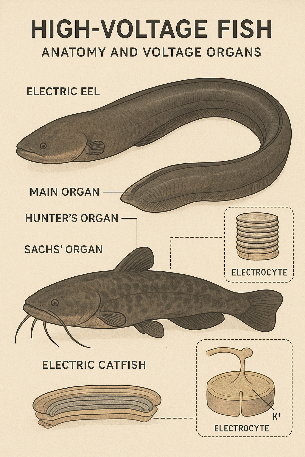
African Electric Catfish (Malapterurus electricus)
- Voltage output: Up to 450 volts (typically 300-400 volts)
- While not consistently exceeding 600 volts, exceptional specimens have been reported
- Distribution: Throughout tropical Africa
- Maximum length: 1.2 meters
- Unique among electric fish for having a single, continuous electric organ
Pacific Electric Ray (Torpedo californica)
- Voltage output: Up to 50 volts (most rays produce much less)
- Note: While electric rays were historically reported to produce higher voltages, modern measurements confirm most species produce under 100 volts
- The ancient reports of 200+ volts may have reflected cumulative effects or measurement errors
It’s important to note that the Electrophorus genus stands alone in reliably producing voltages exceeding 600 volts, making them the primary focus of high-voltage electric fish research.
Evolutionary Origins of Electrogenesis
Convergent Evolution
One of the most remarkable aspects of electric fish is that electrogenesis evolved independently at least six times in the history of fish evolution. This convergent evolution occurred in:
- South American knifefishes (Gymnotiformes)
- African elephantfishes (Mormyroidea)
- Electric rays (Torpediniformes)
- Electric catfishes (Malapteruridae)
- African electric rays (Narcinidae)
- Stargazers (Uranoscopidae)
Each lineage developed electric organs from different tissue origins, yet arrived at remarkably similar cellular mechanisms—a testament to the power of natural selection.
From Muscle to Electric Organ
The evolutionary pathway from ordinary muscle cells to specialized electrocytes (electric cells) has been reconstructed through comparative anatomy and molecular biology. The process involved:
- Gene Duplication and Modification: Sodium channel genes, crucial for muscle contraction, were duplicated and modified. These changes allowed for faster discharge and recovery cycles.
- Loss of Contractile Function: Muscles that would become electric organs gradually lost their contractile proteins while enhancing their electrical properties.
- Cellular Reorganization: Cells became flattened and stacked like batteries in series, with synchronized neural control.
- Innervation Specialization: Nervous system connections evolved to trigger millions of electrocytes simultaneously, creating a synchronized discharge.
- Morphological Transformation: What began as swimming muscles transformed into dedicated electric organs occupying much of the fish’s body.
The Sodium Channel Revolution
Recent research has revealed that changes in voltage-gated sodium channels were crucial to the evolution of high-voltage discharge. In Electrophorus, specific mutations in the SCN4A gene allowed electrocytes to fire more rapidly and recover faster, enabling the rapid, powerful discharges characteristic of these fish.
Anatomy of High-Voltage Electric Fish
External Morphology of Electrophorus
Electric eels possess a distinctive elongated body that immediately sets them apart from true eels:
Body Plan
- Elongated, cylindrical form tapering toward the tail
- Length: 1.2 to 2.5 meters depending on species
- Weight: Up to 20 kilograms
- Coloration: Dark gray to brown dorsally, yellow-orange ventrally
- Skin: Scaleless, covered with thick mucus layer for insulation
Head Structure
- Flattened, somewhat squared profile
- Small eyes with limited visual acuity
- Large mouth with conical teeth (unlike true eels)
- Gill openings positioned on the throat (ventral surface)
- Specialized sensory pores for electroreception
Fins and Locomotion
- Greatly elongated anal fin running from just behind the head to the tail tip
- Contains 300+ fin rays
- Primary means of locomotion through undulating waves
- Pectoral fins present but small
- Dorsal fin absent
- Caudal (tail) fin reduced or absent
- This locomotion system allows forward and backward movement with equal facility
Internal Anatomy: The Electric Organ System
The most remarkable feature of electric eels is the presence of three specialized electric organs that occupy approximately 80% of the body cavity:
1. Main Electric Organ (Hunter’s Organ)
Location and Structure
- Occupies the ventral portion of the posterior 2/3 of the body
- Largest of the three organs
- Contains approximately 5,000-6,000 electrocytes arranged in 70-100 columns
- Each electrocyte is a modified muscle cell shaped like a flattened disc
Cellular Architecture The electrocytes in the main organ are arranged in a precise geometry:
- Stacked in series like batteries in a flashlight
- Each cell is 0.1-0.15 mm thick
- Separated by connective tissue and innervated on one surface
- One surface (posterior) is densely packed with sodium channels
- The other surface (anterior) has potassium channels and pumps
Function
- Primary weapon for predation and defense
- Produces the characteristic high-voltage discharge
- Can generate 600-860 volts depending on species and individual size
- Discharge duration: 2-3 milliseconds
- Can discharge 400+ times per hour without exhaustion
2. Sach’s Organ
Location and Structure
- Located dorsally and anteriorly to the main organ
- Smaller than the main organ, containing fewer electrocytes
- Approximately 1,500-2,000 electrocytes
- Similar cellular structure to main organ but less densely packed
Function
- Produces lower voltage pulses (10-50 volts)
- Used for navigation and electrolocation
- Emits continuous low-voltage pulses to sense the environment
- Can detect impedance changes caused by objects, prey, or predators
- Operating frequency: 10-25 Hz during rest, up to 50 Hz when active
3. Hunter’s Organ (Posterior Electric Organ)
Location and Structure
- Most posterior of the three organs
- Smallest electric organ
- Contains approximately 500-1,000 electrocytes
- Cellular structure similar to Sach’s organ
Function
- Also produces low-voltage discharges
- Works in concert with Sach’s organ for electrolocation
- Contributes to communication between individuals
- May help coordinate with main organ during high-voltage attacks
The Electrocyte: Nature’s Battery Cell
The electrocyte represents one of evolution’s most elegant solutions to energy storage and rapid release. Understanding its structure and function is key to comprehending high-voltage discharge:
Cellular Morphology
- Disc-shaped, 15-100 micrometers thick
- Diameter: 2-3 millimeters
- Two distinct faces: innervated (posterior) and non-innervated (anterior)
- Extensively folded membranes increase surface area
Membrane Specialization
Posterior Face (Innervated)
- Densely packed with voltage-gated sodium channels (up to 6,000 channels per square micrometer)
- Contains nicotinic acetylcholine receptors
- Receives direct innervation from spinal electromotor neurons
- Capable of rapid depolarization when stimulated
Anterior Face (Non-innervated)
- Contains potassium channels and sodium-potassium pumps
- Maintains resting membrane potential
- Does not participate in the action potential
- Creates electrical asymmetry essential for voltage generation
Resting State
- Maintains a resting potential of approximately -85 millivolts
- Sodium-potassium pumps actively maintain ion gradients
- Interior negative relative to exterior
- ATP consumption even at rest
Discharge Mechanism When triggered by neural command:
- Acetylcholine released at neuromuscular-like junctions on posterior face
- Sodium channels open almost instantaneously
- Sodium ions rush into the cell through posterior membrane
- Posterior face potential reverses: -85 mV becomes +65 mV
- Anterior face remains at resting potential
- Net potential difference across cell: 150 mV
- Thousands of cells in series multiply this voltage
- Total discharge: voltage = (number of electrocytes) × (150 mV)
Recovery Phase
- Sodium channels inactivate within milliseconds
- Potassium channels open, repolarizing the cell
- Sodium-potassium pumps restore ion gradients
- Entire cycle completed in 3-5 milliseconds
- Cell ready to discharge again within 10-20 milliseconds
Neural Control Systems
The synchronized discharge of thousands of electrocytes requires sophisticated neural coordination:
Electromotor Neurons
- Specialized spinal motor neurons
- Large diameter axons for rapid conduction
- Minimal synaptic delay
- Myelinated for maximum conduction velocity
Command Nucleus
- Located in the medulla oblongata
- Coordinates voluntary discharge
- Integrates sensory information
- Can adjust discharge pattern based on circumstance
Synchronization Mechanism
- All electromotor neurons fire within 100 microseconds of each other
- Achieved through:
- Electrical coupling between neurons
- Specialized fast-conducting pathways
- Minimal variation in axon length to different regions
- Equidistant innervation patterns
Respiratory Adaptations
Electric eels face a unique challenge: their electric organs occupy space normally reserved for internal organs, particularly swimming muscles and gills.
Obligate Air-Breathing
- Must surface every 10-15 minutes to breathe air
- Highly vascularized oral cavity acts as accessory breathing organ
- Can extract up to 76% of oxygen from gulped air
- Allows survival in oxygen-poor Amazonian waters
Modified Gills
- Reduced gill surface area compared to similar-sized fish
- Provide only 20-30% of oxygen requirements
- Primarily serve osmoregulatory function
- Compensated by air-breathing capability
Circulatory Adaptations
High Metabolic Demands
- Electric organs consume enormous energy
- Heart rate increases during active discharge
- Extensive capillary networks throughout electric organs
- Blood flow can be redirected to electric organs during active periods
Specialized Circulation
- Multiple arterial supplies to electric organs
- Venous drainage designed to remove metabolic waste quickly
- Counter-current exchange systems maintain temperature
- Prevents overheating during extended discharge sequences
Digestive System Modifications
Despite occupying 80% of body volume with electric organs, electric eels maintain functional digestive systems:
Compressed Anatomy
- Stomach, liver, and intestines compressed into anterior 20% of body
- Highly coiled intestine maximizes surface area
- Large liver stores energy as glycogen and fat
- Can consume prey up to 30% of body length
Feeding Mechanism
- Carnivorous, feeding on fish, amphibians, birds, and small mammals
- Uses electric discharge to stun prey before swallowing
- Powerful suction feeding
- Teeth designed for gripping, not cutting
- Swallows prey whole
Sensory Systems
Electroreception
- Ampullae of Lorenzini-like structures detect electrical fields
- Can sense voltage gradients as small as 10 microvolts/cm
- Used to detect prey buried in substrate
- Essential for navigation in murky water
Lateral Line System
- Detects water movement and vibration
- Supplements electroreception
- Hair cells respond to pressure waves
- Helps locate surface for breathing
Vision
- Small eyes with limited acuity
- Adapted for low-light conditions
- Secondary to electrical sense
- Primarily detects movement and silhouettes
Chemoreception
- Olfactory organs detect chemical cues
- Important for finding carrion
- May play role in reproduction
- Compensates for limited vision
Skeletal and Muscular Adaptations
Axial Skeleton
- Vertebral column contains 100-120 vertebrae
- Elongated compared to body width
- Provides framework for electric organs
- Flexible to allow undulating swimming
Reduced Musculature
- Swimming muscles greatly reduced
- Concentrated around anal fin
- Compensated by efficient undulation
- Energy redirected to electric organ function
Connective Tissue Framework
- Extensive collagen sheets separate electrocyte columns
- Provides electrical insulation between columns
- Maintains structural integrity
- Allows independent column function
Integumentary System
Skin Structure
- Thick epidermis with mucus-secreting cells
- Provides electrical insulation
- Reduces water resistance
- Protects against pathogens
Mucus Functions
- Electrical insulation prevents self-stunning
- Reduces friction during swimming
- Antimicrobial properties
- May play role in communication
Reproductive Anatomy
Sexual Dimorphism
- Females generally larger than males
- Males develop breeding tubercles during spawning
- Internal anatomy similar between sexes
Reproductive Organs
- Gonads occupy limited space anterior to electric organs
- Ovaries can contain thousands of eggs
- Testes produce milt seasonally
- Reproductive cycle tied to wet season
Physiological Mechanisms of High-Voltage Discharge
Bioelectrical Principles
The production of high voltage in living tissue seems paradoxical given that individual cells can only generate about 150 millivolts. Electric eels overcome this limitation through elegant biophysical principles:
Series Arrangement
- Electrocytes stacked like batteries in series
- Voltages sum across cells
- 5,000 cells × 0.15 V = 750 V potential
- Insulating tissues prevent short-circuits
Parallel Arrangement
- Multiple columns discharge simultaneously
- Increases current capacity (amperage)
- Typical discharge: 1 ampere
- Power output: 600-860 watts for 2 milliseconds
Energy Budget
The metabolic cost of maintaining and operating electric organs is substantial:
Basal Maintenance
- Sodium-potassium pumps consume ATP continuously
- Approximately 20% of resting metabolic rate
- Higher in larger individuals with more electrocytes
- Requires constant food intake
Discharge Energy
- Single high-voltage discharge: ~0.3-0.5 joules
- Can discharge 400+ times per hour
- Total energy expenditure: 120-200 joules per hour during active hunting
- Recovered through increased feeding and metabolic rate
Thermal Management
Repeated discharge generates heat:
Heat Production
- Each discharge produces waste heat
- Concentrated in electric organs
- Can raise tissue temperature 2-3°C during sustained activity
- Dissipated through circulation and skin
Cooling Mechanisms
- Increased blood flow to electric organs
- Behavioral thermoregulation (moving to cooler water)
- Large surface area to volume ratio aids cooling
- Mucus layer facilitates heat transfer to water
Ecological Role and Behavior
Predatory Behavior
Electric eels employ sophisticated hunting strategies:
Prey Detection
- Low-voltage pulses detect prey through electroreception
- Can sense hidden fish, buried invertebrates
- Effective range: 3-5 meters in optimal conditions
- Works in complete darkness
Attack Sequence
- Detection through electrolocation
- Approach using undulating swimming
- Position body to optimize discharge effectiveness
- Deliver multiple high-voltage pulses (3-5 rapid bursts)
- Prey immobilized through involuntary muscle contractions
- Consumption while prey stunned
Induced Muscle Spasm Recent research has shown that electric eel discharges don’t just stun—they remotely control prey muscles:
- Discharge activates prey motor neurons
- Causes violent whole-body muscle contractions
- Prevents escape and reveals hidden prey
- More effective than simple paralysis
Defense Mechanisms
Anti-Predator Strategies
- High-voltage discharge effective against caimans, jaguars, and humans
- Can deliver multiple discharges in rapid succession
- Leaping attack behavior when threatened at surface
- Size serves as additional deterrent
Electrical Display
- May use sub-lethal shocks as warning
- Progressive escalation from low to high voltage
- Conserves energy compared to maximum discharge
Social Behavior
Communication
- Low-voltage organ discharges serve communicative functions
- Each individual has distinctive pulse pattern
- May signal dominance, reproductive state, or alarm
- Electric organ discharge (EOD) varies with behavioral context
Territoriality
- Large individuals maintain territories
- Electrical signals may demarcate boundaries
- Aggressive interactions involve electrical displays
- Subordinate fish alter EOD patterns in presence of dominants
Reproduction and Development
Breeding Behavior
- Seasonal reproduction tied to onset of wet season
- Males construct foam nests in shallow water
- Courtship involves low-voltage electrical exchanges
- Females deposit thousands of eggs in nest
Parental Care
- Males guard eggs and newly hatched larvae
- Use low-voltage pulses to maintain oxygen circulation
- Protect offspring for several weeks
- Young disperse once capable of independent feeding
Development of Electric Organs
- Larvae hatch with non-functional electric organs
- Electrocytes develop from myotomal muscle precursors
- Low-voltage capability develops first (weeks)
- High-voltage capability emerges at 10-15 cm length
- Full adult voltage achieved at sexual maturity
Biomedical and Technological Significance
Medical Research Applications
Neurological Research
- Electric eel electrocytes served as model for understanding action potentials
- Led to voltage clamp technique development
- Contributed to understanding of ion channel diseases
- Inspiration for cardiac pacemaker technology
Acetylcholine Receptor Studies
- Electric organ tissue rich source of receptors
- Enabled purification and characterization
- Advanced understanding of neuromuscular junction
- Relevant to myasthenia gravis research
Sodium Channel Research
- Multiple sodium channel isoforms in electric organs
- Model for understanding channelopathies
- Insights into epilepsy mechanisms
- Drug development target for pain management
Bio-Inspired Technology
Power Generation
- Artificial electric organs being developed
- Potential for implantable power sources
- Could eliminate battery replacement surgeries
- Harvest energy from body movements or gradients
Soft Robotics
- Flexible, distributed actuation systems
- Inspired by electric organ architecture
- Potential for underwater robotics
- Biomimetic swimming mechanisms
Sensor Networks
- Distributed sensing inspired by electroreception
- Applications in underwater navigation
- Low-power communication systems
- Environmental monitoring
Conservation Status and Threats
Current Population Status
Electric eels are not currently listed as endangered, but face several threats:
Habitat Degradation
- Deforestation reduces flooded forest habitat
- Agricultural runoff affects water quality
- Dam construction fragments populations
- Mining operations release toxins
Direct Exploitation
- Traditional medicine trade in some regions
- Aquarium trade (though mostly farm-bred now)
- Inadvertent catch in fishing operations
- Indigenous subsistence use (sustainable)
Conservation Challenges
Knowledge Gaps
- Population sizes largely unknown
- Distribution of recently described species unclear
- Genetic diversity not fully assessed
- Climate change impacts unpredictable
Protected Areas
- Many populations occur in protected reserves
- Indigenous territories provide de facto protection
- Monitoring programs limited
- Enforcement challenges in remote areas
Future Research Directions
Unanswered Questions
Evolutionary Biology
- How did voltage evolve to extreme levels?
- What maintains species boundaries in Electrophorus?
- Why do some populations show intermediate voltages?
- What role does sexual selection play?
Physiology
- Maximum theoretical voltage limits
- Metabolic constraints on discharge frequency
- Aging effects on electric organ function
- Individual variation in discharge characteristics
Ecology
- Population dynamics and density
- Trophic role in ecosystem
- Interspecific interactions with other electric fish
- Impact on prey population structure
Technological Frontiers
Biomimetic Applications
- Improved battery technologies
- Self-powered biomedical devices
- Soft robotic actuators
- Underwater communication systems
Synthetic Biology
- Engineering electrocytes in cell culture
- Creating artificial electric organs
- Optimizing voltage output
- Miniaturization for implantable devices
Conclusion
Electric fish capable of generating over 600 volts represent a pinnacle of evolutionary innovation. The Electrophorus genus, particularly E. voltai with its record-breaking 860-volt discharge, demonstrates nature’s capacity to push biological systems to extraordinary extremes. From modified muscle cells to sophisticated electric organs, from simple ion channels to synchronized neural networks, every aspect of their anatomy reflects millions of years of refinement.
These remarkable creatures have contributed immensely to our understanding of bioelectricity, neuroscience, and evolution. They continue to inspire technological innovation while reminding us of the unexplored wonders in Earth’s aquatic ecosystems. As we face unprecedented environmental challenges, protecting the habitats of electric eels and continuing their study becomes increasingly important—not only for conservation but for the insights they offer into biological possibility and human innovation.


Be First to Comment