

A Complete Guide to Human Bioelectricity and Electrical Circulation
1. Introduction: The Body as a Living Circuit
Every heartbeat, every thought, every muscle twitch, and every sensation of heat or pain begins with the same basic event: a tiny electrical signal crossing a cell membrane. The human body is, in a very literal sense, an electrical system. It does not run on copper wires and silicon chips, but on ions, membranes, and voltage gradients that have been refined by roughly four billion years of evolution. This article explores that system in depth, examining how electrical signals are generated, how they travel, what they control, and why understanding them matters for medicine, technology, and everyday health.
The phrase “electrical circulation” is a useful way to think about this system because, much like the circulation of blood, the circulation of electrical signals in the body is constant, distributed, and essential to life. Unlike blood, which moves through a closed network of vessels driven by the heart, electrical signals move through cells and tissues driven by chemical gradients and specialized proteins embedded in cell membranes. The heart itself, remarkably, generates its own rhythmic electrical impulses that in turn drive the circulation of blood, making the cardiovascular and bioelectric systems deeply intertwined.
Bioelectricity is not a metaphor. It is measurable in volts, describable with the same physics used for batteries and circuits, and detectable using electrodes placed on the skin. An electrocardiogram (ECG), an electroencephalogram (EEG), and an electromyogram (EMG) are all, at their core, voltmeters reading the electrical activity of the heart, brain, and muscles respectively. These technologies exist because the body genuinely produces electrical signals large enough and organized enough to be captured from outside the skin.
This guide is organized to build understanding from the ground up. It begins with the single cell and the molecular machinery that makes a resting cell into a tiny charged battery. It then examines the action potential, the fundamental unit of electrical signaling shared by neurons, muscle cells, and the specialized cells of the heart. From there, the discussion expands outward to full organ systems: the nervous system, the brain, the heart, the muscles, and the sensory organs. Later sections cover the medical technologies built on these principles, the disorders that arise when electrical circulation goes wrong, and the emerging frontier of bioelectronic medicine, where engineered devices interface directly with the body’s own electrical language.
Why does this matter? Because nearly every major category of human disease touches bioelectricity in some way. Heart attacks and arrhythmias are electrical failures as much as mechanical ones. Epilepsy is a disorder of electrical over-excitability in the brain. Diabetic neuropathy, multiple sclerosis, and many muscular disorders involve the breakdown of the electrical insulation and signaling pathways described in this article. Even mental health and cognitive performance are tied to the electrical rhythms of the brain. A working knowledge of the body’s electrical circulation is therefore not a niche curiosity; it is foundational to understanding modern medicine, biomedical engineering, and human physiology as a whole.
2. Historical Foundations: Discovering Animal Electricity
The idea that living tissue could generate and respond to electricity emerged gradually over the eighteenth century, at a time when electricity itself was still a laboratory curiosity produced by friction machines and Leyden jars. The pivotal moment is usually credited to the Italian physician and physicist Luigi Galvani, who in the 1780s observed that the severed legs of dead frogs twitched violently when touched by two different metals connected in a circuit. Galvani concluded that the muscle itself contained an intrinsic “animal electricity,” a vital force distinct from the electricity produced by machines.
Galvani’s contemporary and rival, Alessandro Volta, disagreed. Volta argued that the electricity was not generated by the animal tissue at all, but by the contact between the two dissimilar metals, with the frog’s leg acting merely as a sensitive detector, not a source. To prove his point, Volta built a device made entirely of metal discs and moistened cardboard, with no biological tissue at all, and showed it produced a continuous electric current. This device, first demonstrated around 1800, became known as the voltaic pile, and it is recognized as the first true electric battery in history.
The historical irony is that both men were partly right. Volta was correct that dissimilar metals in contact can generate a current, and his invention of the battery revolutionized physics and enabled the entire subsequent development of electrical science. But Galvani was also correct in a deeper sense: living tissue genuinely does generate its own electricity, entirely independent of any metal contact. Later experiments, particularly those of the German physiologist Emil du Bois-Reymond in the mid-1800s, confirmed that nerves and muscles produce measurable electrical currents on their own, using sensitive galvanometers rather than metal-triggered twitches. Du Bois-Reymond is often regarded as the founder of electrophysiology as a rigorous experimental discipline.
Over the following century, the tools for measuring bioelectricity steadily improved. In 1887, the British physiologist Augustus Waller recorded what is considered the first human electrocardiogram using a capillary electrometer, demonstrating that the electrical activity of the human heart could be captured from the surface of the body. The Dutch physiologist Willem Einthoven refined this technique dramatically in the early twentieth century using the string galvanometer, a highly sensitive instrument that produced clear, reproducible ECG tracings. Einthoven’s system of labeling the ECG waveform with the letters P, Q, R, S, and T is still used universally today, and he received the Nobel Prize in Physiology or Medicine in 1924 for this work.
Electrophysiology of the nervous system advanced in parallel. In the 1930s and 1940s, Alan Hodgkin and Andrew Huxley used the exceptionally large nerve fibers of the squid, known as giant axons, to directly measure the voltage changes occurring during a nerve impulse. Their quantitative model of the action potential, published in 1952, remains the mathematical foundation of neuroscience and earned them the Nobel Prize in 1963, shared with John Eccles. Around the same time, Hans Berger in Germany recorded the first human electroencephalogram in 1924, establishing that the brain itself produces rhythmic electrical oscillations detectable at the scalp.
These historical threads, spanning frog legs, metal piles, string galvanometers, and squid axons, converge on a single insight that took roughly a century and a half to fully establish: living cells generate, store, and transmit electrical energy using specialized biological machinery, and this machinery can be measured, modeled, and eventually engineered. Everything discussed in the remainder of this article rests on that foundation.
3. The Cellular Battery: Membrane Potential and Ion Gradients
To understand electrical circulation in the body, it is necessary to start at the smallest possible scale: the individual cell. Every living cell in the human body, from a skin cell to a neuron, maintains a voltage difference across its outer membrane. This voltage, called the resting membrane potential, typically measures around negative 70 millivolts in a neuron, meaning the inside of the cell is more negatively charged than the outside. This may seem like an insignificant number compared to household electricity, but because the cell membrane is only about seven nanometers thick, this voltage difference corresponds to an electric field of roughly ten million volts per meter, comparable in intensity to a lightning bolt, concentrated across an extraordinarily thin insulating layer.
3.1 The Cell Membrane as an Insulator
The cell membrane is composed of a lipid bilayer, a double layer of fat molecules that is essentially impermeable to charged particles such as ions. This property makes the membrane function like the insulating material in a capacitor, a component that stores electrical charge. On either side of this insulating layer sit conductive fluids, the cytoplasm inside the cell and the extracellular fluid outside, both rich in dissolved ions. The membrane, sandwiched between two conductive fluids, is the physical basis of the cell’s ability to store a voltage difference.
3.2 The Key Players: Sodium, Potassium, Calcium, and Chloride
Four ions dominate the electrical life of the cell: sodium (Na+), potassium (K+), calcium (Ca2+), and chloride (Cl-). Their concentrations differ dramatically between the inside and outside of the cell. Sodium is far more concentrated outside the cell than inside. Potassium is far more concentrated inside the cell than outside. Calcium is kept at extremely low concentration inside the resting cell compared to outside, and chloride is generally more concentrated outside than inside. These concentration differences are not accidents; they are actively maintained by the cell at significant metabolic cost, because the gradients themselves are the stored energy that powers electrical signaling.
3.3 The Sodium-Potassium Pump
The single most important protein responsible for maintaining these gradients is the sodium-potassium ATPase, often called the sodium-potassium pump. This protein sits in the cell membrane and uses the energy from ATP, the cell’s universal energy currency, to move three sodium ions out of the cell for every two potassium ions it moves in, against both of their concentration gradients. Because this pump moves unequal numbers of positive charges across the membrane, it directly contributes a small amount to the resting negative voltage inside the cell, in addition to its larger role of maintaining the concentration gradients that other channels later exploit.
It has been estimated that the sodium-potassium pump consumes a substantial fraction of the resting energy budget of the brain, sometimes cited at somewhere between one third and one half of the brain’s total energy consumption, underscoring just how costly it is for the nervous system to keep its cells electrically charged and ready to fire at every moment.
3.4 Ion Channels and the Nernst Equation
While pumps actively create ion gradients, ion channels are the passive gates that allow ions to flow down those gradients when opened. At rest, the neuron membrane is far more permeable to potassium than to sodium, because a population of “leak” potassium channels remains open even when the cell is not being stimulated. Because potassium tends to flow out of the cell along its concentration gradient, and because this flow leaves the negatively charged proteins inside the cell relatively uncompensated by positive charge, the inside of the cell settles into a negative resting voltage.
The theoretical voltage at which a particular ion would be in perfect equilibrium, with no net flow in or out despite the membrane being permeable to it, can be calculated using an equation known as the Nernst equation. For potassium, this equilibrium potential typically falls close to the actual measured resting potential of most neurons, which is one of the strongest pieces of evidence that resting potential is dominated by potassium permeability. When multiple ion permeabilities are considered together, a related and more general formula called the Goldman-Hodgkin-Katz equation is used to predict the resting membrane potential from the combined effect of sodium, potassium, and chloride permeabilities.
3.5 The Cell as a Charged Battery
Taken together, this arrangement, a thin insulating membrane, unequal ion concentrations on either side, active pumps that maintain those concentrations, and passive channels that allow controlled current flow, makes every living cell a microscopic battery. The chemical energy stored in the ion gradients is the “fuel,” the membrane is the insulator that keeps the charge separated, and the channels are the switches that, when opened, allow that stored energy to be released as an electrical signal. This is the essential machinery from which the entire electrical circulation system of the human body is built.
4. The Action Potential: The Body’s Basic Electrical Signal
If the resting membrane potential is the charged state of the cellular battery, the action potential is the discharge event, a rapid, self-propagating spike of voltage that serves as the fundamental signal of the nervous system, the muscular system, and the heart. Despite the enormous diversity of tissues that use them, action potentials in the human body share a remarkably consistent underlying mechanism, first fully described mathematically by Hodgkin and Huxley using the squid giant axon.
4.1 Threshold and the All-or-Nothing Principle
An action potential does not occur gradually; it either happens completely or not at all, a property known as the all-or-nothing principle. A stimulus must depolarize the membrane, meaning make the inside less negative, past a certain threshold voltage, often around negative 55 millivolts in a typical neuron, in order to trigger the full event. Small stimuli that do not reach threshold simply fade away without producing a spike. Once threshold is crossed, however, the cell does not produce a partial or proportional response; it fires a full, stereotyped spike regardless of how far above threshold the triggering stimulus was.
4.2 Depolarization: The Rising Phase
The rising phase of the action potential is driven by voltage-gated sodium channels, proteins that remain closed at resting potential but snap open when the membrane depolarizes past threshold. Because sodium is far more concentrated outside the cell, and the inside is negatively charged relative to outside, both the concentration gradient and the electrical gradient push sodium ions into the cell once these channels open. This sudden influx of positive charge rapidly drives the membrane potential from its negative resting value toward a positive value, often peaking around positive 30 to 40 millivolts in a neuron, in a process that takes less than a millisecond.
4.3 Repolarization: The Falling Phase
Two mechanisms bring the membrane potential back down. First, the voltage-gated sodium channels do not stay open; after opening, they rapidly inactivate on their own, shutting off further sodium entry regardless of the membrane voltage. Second, voltage-gated potassium channels, which open more slowly than sodium channels, become active during this phase and allow potassium to flow out of the cell along its concentration gradient. This outward flow of positive charge drives the membrane potential back toward, and often briefly below, its resting value, a phase known as hyperpolarization or the undershoot.
4.4 The Refractory Period
Immediately after an action potential, the cell passes through a refractory period during which it is difficult or impossible to trigger another spike. During the absolute refractory period, the sodium channels remain inactivated and no stimulus, however strong, can trigger a new action potential. During the following relative refractory period, sodium channels are gradually recovering and potassium channels may still be open, so a new action potential can be triggered, but only by a stronger than normal stimulus. The refractory period serves two crucial functions: it enforces a maximum firing rate on the cell, and, in the context of a nerve fiber, it ensures that the action potential travels in only one direction rather than reversing course.
4.5 Propagation Along a Fiber
An action potential generated at one point on a nerve or muscle fiber does not stay localized; it triggers a chain reaction. The local depolarization spreads passively to the immediately adjacent membrane, bringing that neighboring patch to its own threshold and triggering a fresh, full-strength action potential there. This process repeats down the length of the fiber, meaning the signal is regenerated at every point rather than simply diffusing and weakening, which is why nerve signals can travel long distances, in some human nerves over a meter, without losing strength.
4.6 Why the Same Mechanism Serves Many Purposes
The remarkable versatility of the action potential is that this same basic electrochemical event, sodium in, potassium out, threshold, refractory period, underlies nerve impulses carrying sensory information from the fingertip, motor commands driving a muscle contraction, and the spreading wave of excitation that triggers each heartbeat. The differences between these systems arise not from a different fundamental mechanism, but from differences in the specific ion channel types involved, the shape and length of the cell, the presence or absence of insulation, and the way individual cells are connected to their neighbors, topics explored in the following sections.
5. Neurons and the Nervous System: Wiring the Body
The nervous system is the body’s primary long-distance electrical communication network, connecting the brain and spinal cord to every muscle, gland, and sensory receptor. Its basic functional unit is the neuron, a specialized cell built almost entirely around the task of receiving, integrating, and transmitting electrical signals.
5.1 The Structure of a Neuron
A typical neuron has three main structural regions. The dendrites are branching extensions that receive incoming signals from other neurons, typically in the form of small graded voltage changes. The cell body, or soma, integrates these incoming signals and contains the machinery for maintaining the cell’s general health. The axon is a single, often long, cable-like extension that carries the outgoing action potential away from the cell body toward its target, whether that target is another neuron, a muscle fiber, or a gland. At the far end of the axon, the signal reaches specialized junctions called synapses, where it is transmitted to the next cell.
5.2 The Synapse: Electrical Signal Becomes Chemical Signal
At most synapses in the human nervous system, the transmission of the signal from one neuron to the next is not purely electrical but chemical. When an action potential reaches the end of the axon, called the axon terminal, it triggers voltage-gated calcium channels to open. The resulting influx of calcium causes small membrane-bound sacs called synaptic vesicles, each containing a chemical neurotransmitter, to fuse with the cell membrane and release their contents into the narrow gap between the two neurons, known as the synaptic cleft. The neurotransmitter then diffuses across this gap and binds to receptor proteins on the receiving neuron, which in turn opens or closes ion channels on that neuron, producing a new, smaller electrical signal.
This electrical-to-chemical-to-electrical relay might seem like an inefficient extra step, but it provides tremendous computational flexibility. Because different neurotransmitters and receptor combinations can either excite or inhibit the receiving neuron, and because a single neuron may receive thousands of synaptic inputs from different sources simultaneously, the chemical synapse allows the nervous system to perform complex integration, essentially a form of biological computation, before deciding whether to generate a new action potential. Some specialized synapses in the human body, called electrical synapses or gap junctions, skip the chemical step entirely and allow the electrical current itself to pass directly between two cells, enabling extremely fast and highly synchronized signaling in certain tissues.
5.3 Myelin and Saltatory Conduction
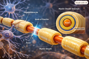
Many axons in the human body, particularly those responsible for fast reflexes and voluntary movement, are wrapped in a fatty insulating substance called myelin, produced by specialized support cells known as Schwann cells in the peripheral nervous system and oligodendrocytes in the brain and spinal cord. Myelin is not a continuous sheath; it is interrupted at regular intervals by small unmyelinated gaps called nodes of Ranvier.
Because the myelinated segments act as excellent electrical insulation, voltage-gated sodium channels are concentrated almost exclusively at the nodes of Ranvier rather than being spread evenly along the axon. The action potential effectively jumps from node to node, a process called saltatory conduction, from the Latin word for “leaping.” This dramatically increases conduction speed while also conserving metabolic energy, since the cell only needs to regenerate the action potential at the sparse nodes rather than continuously along the entire length of the fiber. The fastest myelinated nerve fibers in the human body can conduct signals at speeds exceeding 100 meters per second, while unmyelinated fibers, which carry signals such as dull, chronic pain, may conduct at less than two meters per second.
5.4 The Peripheral and Central Nervous Systems
The nervous system is broadly divided into the central nervous system, comprising the brain and spinal cord, and the peripheral nervous system, comprising all the nerves that branch out to the rest of the body. The peripheral nervous system is further divided into the somatic nervous system, which controls voluntary movement and carries conscious sensory information, and the autonomic nervous system, which regulates involuntary functions such as heart rate, digestion, and pupil dilation through its two opposing branches, the sympathetic and parasympathetic systems. All of these divisions rely on the same fundamental electrical machinery described above, organized into vast, specialized networks that together allow the body to sense its environment, coordinate movement, and regulate its internal state.
6. The Brain’s Electrical Networks
The human brain contains on the order of 86 billion neurons, each connected to thousands of others, forming a network whose combined electrical activity is complex enough that it is often described using the language of orchestras and symphonies rather than simple circuits. Yet at its foundation, brain activity is still built from the same action potentials and synaptic transmissions described in the previous sections, simply organized on an almost unimaginable scale.
6.1 Neural Oscillations and Brainwaves
When large populations of neurons fire in a coordinated, rhythmic fashion, their combined electrical activity produces oscillations that can be detected from outside the skull using an electroencephalogram. These oscillations are conventionally grouped into frequency bands, each loosely associated with particular states of arousal or cognitive activity. Delta waves, the slowest, are most prominent during deep, dreamless sleep. Theta waves are associated with drowsiness, meditation, and certain memory processes. Alpha waves typically dominate when a person is calm and relaxed with eyes closed. Beta waves are associated with active concentration and normal waking alertness. Gamma waves, the fastest commonly studied band, are linked to high-level cognitive processing and the binding together of information from different brain regions into a unified perception.
6.2 Why the Brain Oscillates
Neural oscillations are not simply background noise; they appear to serve as an organizing mechanism that allows distant brain regions to coordinate their activity. When two populations of neurons oscillate in synchrony, they are more likely to influence each other effectively, because their moments of peak excitability line up in time. This synchronization is thought to underlie processes such as attention, working memory, and the integration of sensory information from different modalities into a single coherent experience.
6.3 The Electroencephalogram in Practice
An EEG is recorded by placing an array of electrodes on the scalp, typically arranged according to a standardized layout, and measuring the tiny voltage differences, usually in the range of microvolts, between pairs of electrodes over time. Because the skull and scalp act as resistive and somewhat distorting layers between the brain and the electrode, the EEG signal represents the summed and blurred activity of millions of neurons rather than the crisp firing of any single cell. Despite this blurring, EEG remains an extraordinarily useful clinical tool because it has excellent temporal resolution, capturing changes in brain activity on the order of milliseconds, which is far faster than imaging techniques based on blood flow.
6.4 Clinical Applications
EEG is the primary diagnostic tool for epilepsy, since it can directly capture the abnormal, hypersynchronous electrical discharges that define a seizure, often distinguishing between different seizure types based on where the discharge originates and how it spreads. EEG is also used to assess sleep architecture in sleep laboratories, to monitor the depth of anesthesia during surgery, to help diagnose certain causes of altered consciousness, and, in cases of severe brain injury, to help determine whether meaningful brain activity is still present.
6.5 Beyond the Scalp: Deeper Recording Techniques
In some clinical and research situations, electrodes are placed directly on or within the surface of the brain itself, techniques known as electrocorticography and intracranial or depth electrode recording respectively. These invasive methods, generally reserved for patients undergoing evaluation for epilepsy surgery or for certain research studies, provide far higher spatial resolution and signal quality than scalp EEG, since they avoid the blurring effect of the skull, and have contributed enormously to modern understanding of how localized brain regions generate and coordinate their electrical activity.
7. The Heart’s Electrical Conduction System
Of all the electrical systems in the human body, the heart’s is perhaps the most elegant, because it must generate a perfectly timed, self-sustaining rhythm without any conscious control, repeating roughly once per second for an entire human lifetime, amounting to more than two and a half billion beats over an average lifespan.
7.1 The Sinoatrial Node: The Heart’s Natural Pacemaker
The heartbeat originates in a small cluster of specialized cells located in the upper wall of the right atrium, called the sinoatrial node, or SA node. Unlike ordinary neurons and muscle cells, which sit quietly at a stable resting potential until stimulated, the cells of the SA node possess an unstable resting potential that drifts slowly upward on its own, a property called automaticity, driven largely by a specialized set of ion channels sometimes referred to informally as “funny” channels because of their unusual behavior. Once this slow drift reaches threshold, the SA node cells fire an action potential spontaneously, without any external nerve input required, at an intrinsic rate of roughly 60 to 100 times per minute in a healthy adult at rest.
7.2 Spreading the Signal: Atria to Ventricles
From the SA node, the electrical impulse spreads outward through the muscle cells of both atria, causing them to contract and push blood into the ventricles. Cardiac muscle cells are electrically connected to one another through specialized low-resistance connections called gap junctions, concentrated at structures called intercalated discs, which allow the wave of depolarization to pass directly from one muscle cell to the next without needing a nerve fiber to relay it. This is why the heart is often described as a functional syncytium, a mass of individual cells that behaves electrically as a single connected unit.
The impulse then reaches a second specialized cluster of cells, the atrioventricular node, or AV node, located at the boundary between the atria and ventricles. The AV node deliberately conducts the signal more slowly than the surrounding tissue, introducing a brief delay of roughly one-tenth of a second. This delay is critically important because it allows the atria to finish contracting and fully empty their blood into the ventricles before the ventricles themselves begin to contract, ensuring the two chambers work in proper sequence rather than simultaneously.
7.3 The Bundle of His and Purkinje Fibers
After passing through the AV node, the signal enters a specialized conduction pathway called the bundle of His, which splits into left and right bundle branches that travel down either side of the wall separating the two ventricles. These bundle branches then fan out into a dense network of rapidly conducting cells called Purkinje fibers, which spread throughout the ventricular walls. Purkinje fibers conduct electrical impulses considerably faster than ordinary cardiac muscle, which allows the depolarization signal to reach nearly all parts of both ventricles almost simultaneously. This near-simultaneous activation is essential for an efficient, coordinated contraction that squeezes blood out of the heart effectively, rather than a disorganized, wave-like contraction that would pump blood poorly.
7.4 Reading the Signal: The Electrocardiogram
The electrocardiogram captures this entire sequence of electrical events from electrodes placed on the skin, producing a characteristic waveform with distinct named components. The P wave represents the depolarization, or electrical activation, of the atria. The QRS complex, a sharp, tall spike, represents the depolarization of the ventricles, and because the ventricular muscle mass is much larger than the atrial mass, this spike is correspondingly much larger in amplitude than the P wave. The T wave represents the repolarization, or electrical recovery, of the ventricles as they reset for the next beat. Notably, atrial repolarization also occurs but produces a wave typically too small to see, since it is masked by the much larger QRS complex occurring at nearly the same time.
The time intervals between these waves are as clinically important as the waves themselves. The PR interval reflects the delay introduced by the AV node, and an abnormally long PR interval can indicate a conduction block at that node. The QT interval reflects the total duration of ventricular depolarization and repolarization combined, and an abnormally prolonged QT interval is associated with an increased risk of dangerous ventricular arrhythmias. Physicians and cardiac technicians are trained to read these intervals and waveform shapes to diagnose an enormous range of cardiac conditions, from simple rhythm disturbances to acute heart attacks, often within minutes of recording the tracing.
7.5 Autonomic Modulation of Heart Rhythm
While the SA node sets the heart’s intrinsic rhythm on its own, this rate is continuously adjusted by the autonomic nervous system. The sympathetic nervous system, associated with the body’s “fight or flight” response, releases signaling molecules that speed up the SA node’s drift toward threshold, increasing heart rate. The parasympathetic nervous system, primarily through the vagus nerve, releases signals that slow this drift, decreasing heart rate. This is why heart rate rises during exercise, stress, or excitement, and falls during rest, relaxation, or sleep, all without any conscious control over the underlying electrical machinery.
8. Muscles and Electrical Activation
Every voluntary and involuntary movement of the human body, from lifting a coffee cup to the peristaltic churning of the intestines, depends on converting an electrical signal into a mechanical force. This conversion process is called excitation-contraction coupling, and it represents one of the most important electrical-to-mechanical transformations in human physiology.
8.1 The Neuromuscular Junction
Skeletal muscles, the muscles attached to bone that produce voluntary movement, are activated by motor neurons whose axons travel from the spinal cord out to the muscle fiber. The specialized synapse between a motor neuron and a muscle fiber is called the neuromuscular junction. When an action potential arrives at this junction, it triggers the release of the neurotransmitter acetylcholine, which binds to receptors on the muscle fiber membrane and opens channels that allow positive ions to flow in, generating a new action potential on the muscle fiber itself, called an end-plate potential once it reaches sufficient size to trigger a full muscle action potential.
8.2 From Membrane to Myofilament: The T-Tubule System
Unlike a neuron, whose action potential is largely confined to a thin membrane, a muscle fiber must transmit its electrical signal deep into the interior of a relatively large cell in order to trigger contraction throughout its full thickness almost instantaneously. This is accomplished through a network of inward folds of the muscle cell membrane called transverse tubules, or T-tubules, which carry the action potential rapidly into the cell’s interior. There, the T-tubules sit adjacent to an internal calcium storage compartment called the sarcoplasmic reticulum. The arrival of the electrical signal at this junction triggers the release of stored calcium ions into the muscle cell’s cytoplasm.
8.3 Calcium: The Link Between Electricity and Movement
The sudden rise in cytoplasmic calcium is the direct trigger for mechanical contraction. Calcium ions bind to a regulatory protein called troponin, which is attached to the thin filaments of the muscle’s contractile apparatus. This binding causes a shape change that moves another protein, tropomyosin, out of the way, exposing binding sites on the thin filament that allow the thick filament protein myosin to attach and pull, using chemical energy from ATP, in a repeated ratcheting motion known as the cross-bridge cycle. This is the fundamental molecular basis of all muscle contraction in the human body, and it is entirely dependent on the preceding electrical signal to release the calcium that makes it possible.
8.4 Cardiac and Smooth Muscle Variations
Cardiac muscle uses a broadly similar excitation-contraction coupling mechanism, but with an important addition: the action potential itself allows a small amount of external calcium to enter the cell through voltage-gated calcium channels, and this incoming calcium triggers the release of a much larger amount of calcium from the sarcoplasmic reticulum, a process called calcium-induced calcium release. Smooth muscle, found in the walls of blood vessels, the digestive tract, and many other internal organs, has yet another variation, often relying more heavily on direct calcium entry and on hormonal or stretch-induced activation rather than fast, discrete action potentials, producing the slower, more sustained contractions characteristic of these tissues.
8.5 The Electromyogram
The electrical activity of skeletal muscle can be recorded using an electromyogram, or EMG, either with surface electrodes placed on the skin or with fine needle electrodes inserted directly into the muscle. EMG recordings are used clinically to distinguish between disorders originating in the muscle itself, disorders of the nerves supplying the muscle, and disorders at the neuromuscular junction, since each of these produces a distinctive electrical signature. EMG is also widely used in research and in the control of advanced prosthetic limbs, where the electrical signals recorded from a person’s remaining muscles can be interpreted by a computer to control the movement of a robotic hand or arm.
9. Bioelectricity in the Senses
Every sense the human body possesses, sight, hearing, touch, taste, smell, and balance, ultimately depends on specialized receptor cells converting a physical or chemical stimulus into an electrical signal that the nervous system can process, a process broadly known as sensory transduction.
9.1 Vision and the Electroretinogram
In the retina at the back of the eye, light-sensitive cells called photoreceptors, comprising rods for low-light vision and cones for color vision, contain pigment molecules that change shape when struck by photons of light. This shape change triggers a cascade of chemical reactions that ultimately closes ion channels in the photoreceptor membrane, an unusual arrangement in which light causes the cell to become more, rather than less, electrically negative inside. This altered electrical signal is then relayed through several layers of retinal neurons before reaching retinal ganglion cells, whose axons form the optic nerve and carry the visual signal, now encoded as a pattern of action potentials, to the brain. The combined electrical response of the retina to a flash of light can be recorded from the surface of the eye using a technique called the electroretinogram, used clinically to diagnose various retinal diseases.
9.2 Hearing and the Cochlea
In the inner ear, sound waves cause fluid within a spiral-shaped structure called the cochlea to vibrate, which in turn causes a thin membrane called the basilar membrane to move. This mechanical movement bends tiny hair-like projections, called stereocilia, on top of specialized sensory cells known as hair cells. Bending these stereocilia physically opens mechanically-gated ion channels at their tips, allowing positive ions to flow into the hair cell and generate an electrical signal, a remarkably direct conversion of mechanical vibration into bioelectricity, occurring on a timescale fast enough to preserve the detailed frequency information needed for speech and music perception.
9.3 Touch, Pain, and Temperature
The skin contains a diverse population of sensory nerve endings, each tuned to detect a different type of stimulus. Specialized mechanoreceptors respond to light touch, pressure, vibration, or skin stretch by opening mechanically-gated ion channels similar in principle to those in the inner ear. Thermoreceptors respond to specific temperature ranges using a family of channels known as transient receptor potential channels, some of which are also activated by certain chemicals, which is why capsaicin, the active compound in chili peppers, produces a genuine sensation of heat by directly activating a heat-sensing channel. Nociceptors, the receptors responsible for pain, respond to potentially damaging mechanical, thermal, or chemical stimuli and, importantly, the sensitivity of these fibers can be altered by inflammation, which is part of why an already-damaged area of skin becomes more sensitive to touch during the healing process.
9.4 The Skin’s Galvanic Response
Beyond sensory transduction, the skin itself exhibits a measurable bioelectric phenomenon known as the galvanic skin response, sometimes called electrodermal activity. This refers to changes in the skin’s electrical conductivity that occur when sweat glands, which are controlled by the sympathetic nervous system, become more active in response to emotional arousal, stress, or attention. Because sweat is an electrolyte-rich fluid, even a small increase in sweat gland activity measurably increases the skin’s ability to conduct electricity. This effect is the physiological basis for polygraph testing and is also widely used in psychological and physiological research as an objective, if imperfect, index of emotional arousal.
10. Bioelectric Signaling Beyond Nerves and Muscles
While the nervous system, heart, and muscles are the most well-known users of bioelectricity, research over the past several decades has revealed that electrical signaling plays important roles in tissues not traditionally considered “electrical” at all, including the process of wound healing and even the earliest stages of embryonic development.
10.1 Endogenous Electric Fields in Wounds
Intact skin maintains a voltage difference across its outer layers, generally on the order of tens of millivolts, maintained by ion pumps in skin cells, with the outer surface typically more negative relative to deeper layers. When the skin is wounded, this insulating barrier is broken, and current is able to flow out of the wound, creating a local electric field within and around the injury that can persist for as long as the wound remains open. Research using sensitive voltage-sensing probes has shown that this naturally occurring wound electric field is not simply a byproduct of injury; the cells responsible for closing the wound, including skin cells called keratinocytes, appear to sense the direction of this field and migrate along it, a phenomenon known as galvanotaxis, contributing to the coordinated, directional healing of the wound.
10.2 Bioelectricity in Embryonic Development
Long before a nervous system exists, developing embryonic cells maintain their own resting membrane potentials and communicate with neighboring cells through gap junctions, and researchers studying developmental biology have found that patterns of voltage across groups of embryonic cells can influence how tissues and organs form. Experimental work, much of it conducted using amphibian and other model organisms, has shown that artificially altering the bioelectric state of certain embryonic cells can influence the development of structures such as limbs and even, in striking experiments, the patterning of structures like eyes forming in unexpected locations, suggesting that bioelectric signals act as an additional layer of instruction alongside the genetic code, helping to coordinate how individual cells organize into complex, correctly shaped tissues and organs.
10.3 Bone Remodeling and Piezoelectricity
Bone tissue exhibits a phenomenon called piezoelectricity, in which mechanical stress on the bone’s collagen fibers generates small electrical charges. It has long been observed clinically that bones remodel themselves in response to the mechanical loads placed upon them, becoming denser along lines of greatest stress, a principle known as Wolff’s Law. The piezoelectric charges generated by mechanical loading are thought to be part of the signaling mechanism that tells bone-building cells, called osteoblasts, and bone-resorbing cells, called osteoclasts, where new bone should be added or removed, linking mechanical stress directly to a bioelectric signal that ultimately shapes the skeleton over a person’s lifetime.
10.4 Why This Matters
This broader view of bioelectricity, extending well beyond the classic domains of nerve and muscle, has significant implications for regenerative medicine. If bioelectric signals genuinely help direct tissue growth, repair, and patterning, then therapies that deliberately manipulate these electric fields, whether through externally applied currents, engineered bioelectric materials, or drugs that target specific ion channels, may eventually offer new ways to accelerate wound healing, improve the success of limb reattachment or regeneration research, and better understand certain birth defects that may arise from disruptions in these early bioelectric patterning signals.
11. Medical Technologies Built on Bioelectricity
Because so much of human physiology is fundamentally electrical, an entire branch of medical technology has developed around measuring, interpreting, and, when necessary, directly intervening in the body’s electrical activity.
11.1 Diagnostic Recording: ECG, EEG, and EMG
The three major diagnostic recording technologies discussed throughout this article, the electrocardiogram, electroencephalogram, and electromyogram, all work on the same basic principle: electrodes placed on the skin detect tiny voltage differences generated by the summed electrical activity of large numbers of cells, and this voltage signal is amplified, filtered, and displayed as a waveform for a clinician to interpret. Modern versions of these technologies are often combined with computerized signal processing that can automatically flag abnormal rhythms, measure precise time intervals, or compare a patient’s recording against large reference databases to assist in diagnosis.
11.2 Cardiac Pacemakers
When the heart’s natural pacemaker or conduction pathways fail to generate or transmit an adequate rhythm, an artificial cardiac pacemaker can be surgically implanted to take over this function. A pacemaker consists of a small battery-powered pulse generator, typically implanted under the skin near the collarbone, connected to one or more thin wires called leads that are threaded through blood vessels into the heart chambers. The device continuously monitors the heart’s own electrical activity and delivers precisely timed electrical pulses only when needed, for instance if the heart rate drops below a programmed threshold, effectively substituting an engineered electrical signal for a failing biological one.
11.3 Defibrillators
Certain dangerous cardiac arrhythmias, most notably ventricular fibrillation, involve the heart’s muscle cells firing chaotically and independently rather than in the coordinated wave described in Section 7, resulting in a heart that quivers ineffectively rather than pumping blood. A defibrillator treats this condition by delivering a brief, strong electrical shock across the chest, which momentarily depolarizes essentially all of the heart’s muscle cells simultaneously. This synchronized reset allows the SA node, if it remains functional, the opportunity to resume control and re-establish a normal, coordinated rhythm. Defibrillators exist both as large hospital devices, as portable automated external defibrillators found in many public spaces, and as small implantable devices, called implantable cardioverter-defibrillators, for patients at high risk of recurrent life-threatening arrhythmias.
11.4 Deep Brain Stimulation and Neuromodulation
Deep brain stimulation involves surgically implanting thin electrodes into specific, carefully targeted regions deep within the brain, connected to a programmable pulse generator implanted elsewhere in the body, similar in concept to a pacemaker but targeting neural rather than cardiac tissue. This technology delivers continuous or patterned electrical pulses that can suppress the abnormal neural activity underlying certain movement disorders, most notably Parkinson’s disease and essential tremor, and is also used, sometimes on an investigational basis, for certain treatment-resistant psychiatric conditions and chronic pain syndromes. Related but less invasive neuromodulation technologies include vagus nerve stimulation and spinal cord stimulation, both of which apply electrical pulses to nerve tissue outside the brain itself to treat conditions ranging from epilepsy to chronic pain.
11.5 Transcutaneous Electrical Nerve Stimulation
Transcutaneous electrical nerve stimulation, commonly abbreviated TENS, is a widely available, non-invasive technology in which mild electrical pulses are delivered through electrodes placed on the skin over a painful area. The precise mechanism is still debated, but leading explanations include the idea that stimulating large, fast-conducting touch and pressure fibers can interfere with the transmission of pain signals carried by smaller, slower fibers at the level of the spinal cord, a concept related to the well-known gate control theory of pain, as well as the possibility that TENS stimulates the release of the body’s own natural pain-relieving chemicals.
11.6 Cochlear Implants and Retinal Prostheses
For individuals with severe hearing loss due to damage to the hair cells of the cochlea, a cochlear implant can restore a functional sense of hearing by bypassing the damaged hair cells entirely. An external microphone and processor convert sound into a pattern of electrical signals, which are transmitted to an array of electrodes surgically placed within the cochlea, directly stimulating the auditory nerve fibers that remain intact. A conceptually similar approach, retinal prostheses, aims to restore partial vision to individuals with certain forms of blindness caused by photoreceptor loss, by using an implanted electrode array to directly stimulate the remaining, healthier layers of retinal neurons, allowing at least some visual information to reach the brain via the optic nerve.
12. When Electrical Circulation Fails: Disorders
Because electrical signaling underlies so much of human physiology, disruptions to this system are responsible for some of the most significant categories of disease encountered in medicine.
12.1 Cardiac Arrhythmias
An arrhythmia is any disturbance in the normal timing, origin, or spread of the heart’s electrical activity. Some arrhythmias, such as atrial fibrillation, arise when the atria are driven by chaotic, disorganized electrical activity rather than a single coordinated wave from the SA node, leading to an irregular and often rapid heartbeat that increases the risk of blood clot formation and stroke. Others, such as heart block, occur when the electrical signal is delayed or fails to pass through the AV node at all, potentially requiring a pacemaker. Ventricular tachycardia and ventricular fibrillation, discussed in the previous section, represent more immediately life-threatening disruptions originating in the ventricles themselves, sometimes triggered by scarred heart tissue following a prior heart attack, which can create abnormal pathways that allow electrical signals to circulate in dangerous, self-sustaining loops.
12.2 Epilepsy
Epilepsy is a neurological condition characterized by a tendency toward recurrent seizures, episodes of abnormal, excessive, and hypersynchronous electrical discharge among populations of neurons in the brain. Depending on where in the brain this abnormal activity originates and how far it spreads, seizures can produce an enormous range of symptoms, from brief lapses in awareness to widespread convulsions involving the entire body. The underlying causes of this electrical over-excitability are diverse, including genetic mutations affecting ion channel proteins, structural brain abnormalities, prior brain injury, and, in many cases, no identifiable cause at all, but the common thread is a breakdown in the normal balance between excitatory and inhibitory electrical signaling in the brain.
12.3 Peripheral Neuropathy
Peripheral neuropathy refers to damage affecting the peripheral nerves, the long fibers that carry electrical signals between the spinal cord and the rest of the body. One of the most common causes worldwide is diabetes mellitus, in which chronically elevated blood glucose levels damage both the small blood vessels that supply nerves and the nerve fibers themselves, along with contributing to progressive damage of the myelin sheath, degrading the fiber’s ability to conduct signals efficiently. This typically produces a characteristic pattern of numbness, tingling, or pain beginning in the feet and hands, the body’s longest nerve fibers, and gradually progressing over time if the underlying blood sugar control is not adequately managed.
12.4 Multiple Sclerosis and Demyelinating Disease
Multiple sclerosis is an autoimmune condition in which the body’s own immune system mistakenly attacks and damages the myelin sheath surrounding nerve fibers in the brain and spinal cord. Without intact myelin, saltatory conduction is disrupted, causing nerve signals to travel more slowly or fail to propagate at all along the affected fibers, which produces the characteristic and highly variable symptoms of the disease, including muscle weakness, sensory disturbances, and visual problems, that depend on precisely which nerve pathways happen to be affected by demyelination at any given time.
12.5 Myasthenia Gravis
Myasthenia gravis is an autoimmune disorder in which the immune system produces antibodies that attack the acetylcholine receptors at the neuromuscular junction described in Section 8. With fewer functional receptors available, the electrical signal from the motor neuron produces a weaker end-plate potential in the muscle fiber, which may fail to reach the threshold needed to trigger a full muscle action potential, particularly with repeated or sustained use. This produces the disease’s hallmark symptom of muscle weakness that characteristically worsens with continued activity and improves somewhat with rest, directly reflecting the underlying failure of the electrical handoff at the neuromuscular junction.
12.6 Long QT Syndrome and Channelopathies
Long QT syndrome is one of a broader category of conditions known as channelopathies, disorders caused by mutations in the genes encoding ion channel proteins themselves. In long QT syndrome, mutations affecting the potassium or sodium channels responsible for cardiac repolarization prolong the QT interval described in Section 7, creating a window of vulnerability during which the heart can be triggered into a dangerous ventricular arrhythmia, sometimes with little or no warning, particularly during exercise, strong emotion, or sudden loud noises depending on the specific genetic subtype involved. Similar channelopathies affecting other tissues can cause certain forms of epilepsy, periodic paralysis, and other disorders, illustrating how a single faulty protein at the molecular level can produce disease at the level of an entire organ system.
13. Measuring the Invisible: Instrumentation and Technique
Detecting the body’s electrical signals from outside the skin is a significant technical challenge, since the voltages involved are extremely small, often in the range of microvolts to millivolts, and must be distinguished from a noisy background of electrical interference.
13.1 Electrodes
The interface between the body’s ionic electrical activity and a recording device’s electronic circuitry is the electrode. Surface electrodes, typically made of silver coated with silver chloride, are placed on the skin, often with a conductive gel to improve electrical contact and reduce the resistance of the skin itself, which can otherwise significantly attenuate the signal. Needle electrodes, used for detailed EMG studies, are inserted directly into muscle tissue to record more localized electrical activity, while intracranial electrodes, used in specialized epilepsy evaluation, are placed on or within the brain itself.
13.2 Amplification and Filtering
Because biological voltage signals are so small, they must be amplified thousands of times before they can be usefully displayed or analyzed, typically using a specialized circuit called a differential amplifier, which amplifies the difference between two electrode sites while rejecting signals that appear identically at both electrodes, a property useful for reducing common sources of electrical interference such as nearby power lines. Recorded signals are also passed through electronic filters that remove frequency components outside the range of interest; for example, EEG recordings typically filter out very slow drifts as well as very fast, high-frequency noise, isolating the biologically meaningful oscillation frequencies described in Section 6.
13.3 Artifact and Noise
A persistent challenge in all bioelectric recording is distinguishing genuine physiological signal from artifact, unwanted electrical interference arising from sources other than the tissue of interest. Common sources of artifact include muscle activity contaminating an EEG recording, eye movements producing large voltage swings that can obscure brain activity, poor electrode contact causing erratic baseline drift, and external electrical equipment introducing interference at the frequency of the local power grid. Skilled clinical technicians and increasingly sophisticated automated software algorithms are both employed to identify and, where possible, remove these artifacts before a recording is interpreted.
13.4 From Analog Signal to Digital Diagnosis
Modern bioelectric recording devices convert the amplified and filtered analog voltage signal into a digital format, sampling the voltage at a high rate, often thousands of times per second for EEG and EMG recordings, and storing the result as a sequence of numbers that can be displayed, stored, and analyzed by computer software. This digitization has enabled powerful automated analysis techniques, including algorithms that can detect abnormal heart rhythms in real time from a wearable monitor, software that can flag suspicious EEG patterns for a neurologist’s review, and machine learning models that are increasingly being explored as tools to detect subtle bioelectric abnormalities that might be difficult for a human observer to identify by eye alone.
14. The Future: Bioelectronics and Neural Interfaces
The historical trajectory described throughout this article, from Galvani’s twitching frog leg to today’s implantable pacemakers and cochlear implants, continues to accelerate, and the frontier of research today lies in creating increasingly sophisticated, high-resolution, and often bidirectional interfaces between engineered electronics and the body’s natural electrical circulation.
14.1 Brain-Computer Interfaces
A brain-computer interface is a system that records electrical activity directly from the brain, whether through scalp electrodes, electrodes placed on the brain’s surface, or electrodes implanted within brain tissue, and translates that activity into commands that can control an external device, such as a computer cursor, a robotic arm, or a communication interface. This technology has already been demonstrated to allow individuals with severe paralysis to control assistive devices using their own neural activity, and ongoing research is focused on increasing the number of electrical signals that can be recorded simultaneously, improving the durability of implanted electrodes over years of use, and developing more sophisticated software to decode the complex, high-dimensional patterns of neural electrical activity into useful commands.
14.2 Closed-Loop Neuromodulation
Traditional neuromodulation devices, such as the deep brain stimulators described in Section 11, generally deliver a continuous or fixed pattern of electrical stimulation regardless of the brain’s current state. Newer closed-loop systems are being designed to continuously monitor the brain’s own electrical activity and adjust stimulation parameters in real time, delivering a pulse of stimulation only when a specific abnormal electrical pattern, such as the onset of a seizure, is detected, in principle allowing for more precise, effective, and energy-efficient treatment than constant, unmodulated stimulation.
14.3 Bioelectronic Medicine
A broader emerging field, sometimes called bioelectronic medicine, is exploring the idea that many conditions traditionally treated with drugs might instead be treated, or treated more precisely, by directly modulating the electrical activity of specific nerves that regulate the affected organ or system. Because the nervous system regulates inflammation, hormone release, and organ function through specific, identifiable electrical pathways, proponents of this approach argue that targeted electrical stimulation of the right nerve, at the right pattern and intensity, could in principle achieve therapeutic effects currently achieved by systemic drugs, potentially with fewer side effects, since the intervention could be confined to a specific electrical pathway rather than circulating throughout the entire body.
14.4 Flexible and Biocompatible Electronics
A significant engineering challenge in all of these applications is that conventional rigid electronic materials are poorly matched to the soft, flexible, and constantly moving tissues of the human body, which can lead to inflammation, scarring, and gradual signal degradation around implanted devices over time. Materials science research is increasingly focused on developing flexible, stretchable, and biocompatible electronic materials, including soft polymer-based electrodes and bioresorbable electronics designed to safely dissolve in the body after their function is no longer needed, aiming to create implantable devices that integrate with living tissue far more gently and durably than earlier generations of rigid metal and silicon-based implants.
14.5 A Convergence of Biology and Engineering
Taken together, these developments point toward a future in which the boundary between biological and engineered electrical systems becomes increasingly blurred, with devices capable not only of listening to the body’s natural bioelectric signals but of speaking back to them in the body’s own electrical language, opening possibilities for treating neurological, cardiac, and other diseases with a precision that would have been unimaginable to the early pioneers of bioelectricity, while also raising important questions about safety, long-term reliability, and the ethical implications of increasingly intimate technological integration with human physiology.
15. Conclusion: Why Electrical Circulation Matters
This article has traced the body’s electrical circulation from its smallest building block, the charged cell membrane maintained by ion pumps and channels, up through the action potential shared by nerves, muscles, and the heart, out to the organized electrical networks of the brain and cardiac conduction system, and finally to the medical technologies and disorders that arise from this remarkable biological machinery.
A few key themes emerge from this survey. First, despite the enormous diversity of tissues and functions involved, bioelectricity in the human body is built from a strikingly small set of shared components: a handful of ion species, a limited family of channel and pump proteins, and the same basic physics of charge separation across an insulating membrane. Second, this shared electrical language allows different organ systems that are structurally very different from one another, a neuron, a cardiac muscle cell, a photoreceptor, a healing skin cell, to nonetheless communicate and coordinate using a common underlying signaling mechanism. Third, and perhaps most importantly for practical purposes, an enormous share of modern medicine, from the routine ECG performed in a doctor’s office to the most advanced implantable neurostimulator, exists specifically because this bioelectric activity is measurable, interpretable, and, in many cases, capable of being restored or corrected when it fails.
Understanding the body’s electrical circulation is therefore valuable not only as an academic exercise in physiology but as a practical foundation for understanding an enormous swath of human health and disease. A person who understands why the heart’s electrical conduction system requires the precise sequence of SA node, AV node, and Purkinje fibers is better equipped to understand why a heart attack or an electrolyte imbalance can produce a dangerous arrhythmia. A person who understands the action potential and the role of myelin is better equipped to understand why diabetes or multiple sclerosis can cause nerve damage, and why that damage produces the particular symptoms it does. And a person who appreciates just how much of modern medical technology, pacemakers, defibrillators, cochlear implants, deep brain stimulators, is fundamentally an engineering response to the body’s own bioelectric signaling is better positioned to understand where the field of medicine is likely to go next, as increasingly sophisticated bioelectronic devices continue to blur the line between biological and engineered electrical circulation.
The human body, in the end, runs on electricity every bit as much as it runs on oxygen and glucose. It is simply a form of electricity generated not by wires and batteries in the conventional sense, but by living cells, ion gradients, and molecular machines refined over the entire history of life on Earth, circulating continuously, silently, and essentially without pause, from the first heartbeat before birth to the last.





Be First to Comment