
Part 1 — Pages 1 to 4
PAGE 1 — INTRODUCTION
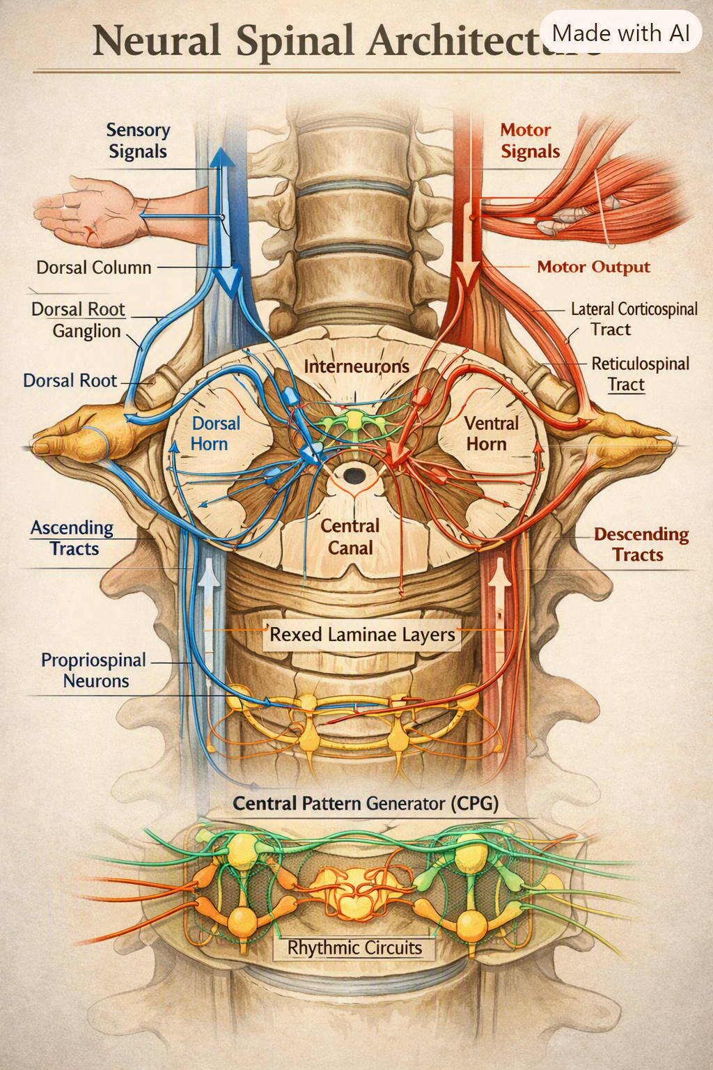
The human spinal cord is a longitudinal neural highway containing millions of neurons arranged in precise architectural layers. It is responsible for transmitting sensory information upward to the brain and motor commands downward to the body. The spinal cord also contains intrinsic neural circuits capable of generating movement patterns without direct brain input — a phenomenon known as central pattern generation (CPG).
Recent research highlights the spinal cord as a dynamic computational organ, not merely a passive conduit. Neurons within the spinal cord exhibit:
- Structured architecture (gray matter horns, white matter tracts)
- Directional movement of signals (afferent vs efferent flow)
- Local circuit integration (interneurons forming micro‑loops)
- Plasticity (synaptic strengthening, rewiring after injury)
Studies from Nature Communications (2026) show that spinal locomotor networks follow spatial and network principles that allow coordinated movement patterns such as walking, running, and balance control.
This thesis explores the full architecture, neuron types, movement pathways, signal propagation, and locomotor circuit dynamics of the spinal cord in deep scientific detail.
PAGE 2 — ARCHITECTURE OF THE SPINAL CORD
2.1 Macro‑Architecture
The spinal cord is organized into two major structural domains:
- Gray Matter (Central “H” Shape) Contains neuronal cell bodies, interneurons, and synaptic networks. Divided into:
- Dorsal Horn — sensory processing
- Lateral Horn — autonomic neurons
- Ventral Horn — motor neurons
- White Matter (Peripheral Columns) Contains ascending and descending myelinated axons. Organized into:
- Dorsal Columns — sensory tracts
- Lateral Columns — mixed sensory/motor tracts
- Ventral Columns — motor tracts
2.2 Micro‑Architecture
Within the gray matter, neurons form laminated layers known as Rexed laminae, each with specialized functions:
- Lamina I–VI: Sensory processing
- Lamina VII: Interneurons and autonomic integration
- Lamina VIII–IX: Motor neuron pools
- Lamina X: Neurons surrounding the central canal
Research from Cell Press (Neuron, 2026) shows that enhancer dynamics regulate the cellular architecture of spinal neurons, influencing differentiation, synaptic patterning, and circuit formation.
2.3 Neuron Types in the Spinal Cord
- Sensory Neurons (Afferent) — enter via dorsal roots
- Motor Neurons (Efferent) — exit via ventral roots
- Interneurons — form local circuits and CPGs
- Autonomic Neurons — regulate visceral organs
- Propriospinal Neurons — connect distant spinal segments
These neurons form vertical and horizontal networks, enabling both segmental and whole‑body coordination.
PAGE 3 — MOVEMENT OF NEURONS & SIGNAL FLOW
3.1 Afferent (Sensory) Flow
Sensory neurons carry information from the body to the spinal cord:
- Receptors detect stimuli (touch, pain, temperature).
- Signals travel through dorsal root ganglia.
- Axons enter the dorsal horn.
- Interneurons relay signals upward through ascending tracts.
3.2 Efferent (Motor) Flow
Motor neurons carry commands from the spinal cord to muscles:
- Descending signals arrive from the brain.
- Motor neuron cell bodies in the ventral horn generate action potentials.
- Axons exit via ventral roots.
- Signals reach skeletal muscles, causing contraction.
3.3 Interneuron Movement & Circuit Dynamics
Interneurons form microcircuits that regulate:
- Reflex arcs
- Locomotor rhythm
- Muscle coordination
- Postural control
These interneurons create feedforward, feedback, and lateral inhibition loops — similar to cortical microcircuits but optimized for rapid movement.
3.4 Central Pattern Generators (CPGs)
CPGs are spinal circuits capable of generating rhythmic movement without brain input.
Research from ScienceDirect (2025) shows that spinal circuits for skilled locomotion rely on interconnected interneuron clusters that produce stable rhythmic outputs.
CPGs control:
- Walking
- Running
- Swimming motions
- Balance adjustments
They operate through oscillatory interneuron networks, modulated by sensory feedback.
PAGE 4 — SYNAPTIC ORGANIZATION & NEURAL MOVEMENT
4.1 Synaptic Layers
Spinal neurons form synapses in highly organized patterns:
- Dorsal horn synapses — sensory integration
- Intermediate zone synapses — interneuron coordination
- Ventral horn synapses — motor output control
Synaptic density varies by region, with motor pools having large multipolar neurons and dense synaptic inputs.
4.2 Neurotransmitters
Key neurotransmitters include:
- Glutamate — excitatory
- GABA & Glycine — inhibitory
- Acetylcholine — motor neuron output
- Norepinephrine & Serotonin — modulatory (descending pathways)
4.3 Movement of Signals
Signal movement follows:
- Longitudinal pathways (ascending/descending tracts)
- Transverse pathways (interneuronal connections)
- Segmental loops (reflex arcs)
This multi‑directional movement allows the spinal cord to act as a distributed processing network.
4.4 Plasticity & Regeneration
Although limited, spinal neurons exhibit:
- Synaptic remodeling
- Axonal sprouting
- Circuit reorganization after injury
Stem‑cell research shows emerging potential for regenerating spinal neurons, as illustrated in recent imaging studies.
PAGE 5 — PROPRIOSPINAL NETWORKS & LONG‑RANGE CONNECTIVITY
Propriospinal neurons form the internal wiring system of the spinal cord, linking distant segments into unified functional networks. These neurons possess long axons that travel vertically across multiple spinal levels, enabling coordination between:
- Cervical and thoracic segments
- Thoracic and lumbar segments
- Lumbar and sacral locomotor centers
Their architecture is essential for whole‑body movement. For example, walking requires synchronized activation of:
- Cervical circuits (arm swing)
- Lumbar circuits (leg movement)
- Sacral circuits (postural stabilization)
Propriospinal neurons act as bridges, ensuring that rhythmic patterns generated in one region propagate smoothly across the entire spinal axis.
5.1 Structural Organization
Propriospinal neurons are categorized into:
- Short‑range propriospinal neurons (within 1–2 segments)
- Long‑range propriospinal neurons (spanning 5–20 segments)
Their axons travel through the ventrolateral funiculus, forming parallel bundles that resemble multi‑lane neural highways.
5.2 Functional Role
They coordinate:
- Interlimb movement
- Postural adjustments
- Bilateral muscle activation
- Reflex modulation
- Locomotor rhythm propagation
Without propriospinal networks, the spinal cord would behave as isolated segments rather than a unified motor system.
PAGE 6 — REFLEX ARC ARCHITECTURE
Reflex arcs are rapid, involuntary neural circuits that bypass the brain to produce immediate responses. Their architecture is one of the most elegant examples of spinal neural engineering.
6.1 Components of a Reflex Arc
- Sensory receptor
- Afferent neuron
- Interneuron
- Motor neuron
- Effector muscle
This loop can complete in 30–50 milliseconds, far faster than cortical processing.
6.2 Types of Reflexes
- Monosynaptic reflexes (e.g., knee‑jerk)
- Polysynaptic reflexes (withdrawal reflex)
- Crossed‑extensor reflexes (balance maintenance)
6.3 Neural Movement Within Reflex Circuits
Reflex circuits demonstrate bidirectional movement:
- Upward sensory flow
- Lateral interneuron processing
- Downward motor output
Interneurons create inhibitory and excitatory balance, ensuring that reflexes are precise rather than chaotic.
PAGE 7 — ASCENDING TRACTS: SENSORY INFORMATION FLOW
Ascending tracts carry sensory information from the body to the brain. Their architecture is highly organized, with each tract dedicated to specific sensory modalities.
7.1 Major Ascending Tracts
- Dorsal Column–Medial Lemniscal Pathway
- Fine touch
- Vibration
- Proprioception
- Spinothalamic Tract
- Pain
- Temperature
- Crude touch
- Spinocerebellar Tracts
- Unconscious proprioception
- Muscle tension feedback
7.2 Movement of Signals
Signals ascend through:
- First‑order neurons (peripheral → spinal cord)
- Second‑order neurons (spinal cord → brainstem)
- Third‑order neurons (thalamus → cortex)
This vertical movement is topographically mapped, meaning the spinal cord preserves spatial organization of the body.
PAGE 8 — DESCENDING TRACTS: MOTOR COMMAND FLOW
Descending tracts carry motor commands from the brain to the spinal cord.
8.1 Major Descending Tracts
- Corticospinal Tract
- Voluntary movement
- Fine motor control
- Reticulospinal Tract
- Posture
- Locomotor initiation
- Vestibulospinal Tract
- Balance
- Head stabilization
- Rubrospinal Tract
- Upper limb coordination
8.2 Movement of Motor Signals
Motor commands descend through:
- Upper motor neurons (cortex → spinal cord)
- Lower motor neurons (spinal cord → muscles)
The architecture ensures precision, speed, and graded force control.




Be First to Comment