The Architecture of All Major Blood Vessels: Blood Flow, Velocity, and Connections to Human Organs
Author
Abstract
The human circulatory system is a highly organized transport network responsible for delivering oxygen, nutrients, hormones, and immune cells to every tissue while removing metabolic waste products. At the center of this network lies the heart, which serves as a powerful biological pump connected to an extensive system of arteries, arterioles, capillaries, venules, and veins. Blood vessels form a continuous circuit that permits communication and physiological integration among all organs.
This thesis explores the architecture of the major blood vessels of the human body, their anatomical organization, patterns of blood flow, velocity characteristics, regulatory mechanisms, and connections to individual organs. It also examines the relationship between vascular structure and physiological function, highlighting how alterations in blood flow contribute to disease.
Table of Contents
- Introduction
- Overview of the Circulatory System
- Blood Composition and Transport
- Structural Architecture of Blood Vessels
- Hemodynamics and Blood Flow Principles
- The Heart as the Central Pump
- The Aorta
- Coronary Circulation
- Cerebral Circulation
- Carotid Arteries
- Vertebral Arteries
- Subclavian Arteries
- Upper Limb Circulation
- Thoracic Circulation
- Pulmonary Circulation
- Abdominal Aorta
- Hepatic Circulation
- Splenic Circulation
- Gastrointestinal Blood Supply
- Renal Circulation
- Endocrine Organ Blood Supply
- Reproductive Organ Circulation
- Pelvic Circulation
- Lower Limb Circulation
- Venous Circulation
- Portal Venous System
- Lymphatic-Vascular Integration
- Blood Flow Velocity in Different Vessels
- Microcirculation
- Capillary Exchange
- Autoregulation of Organ Blood Flow
- Blood Pressure Regulation
- Neural Control of Vessels
- Hormonal Control of Blood Flow
- Fetal Circulation
- Aging and Vascular Changes
- Vascular Diseases
- Organ-Specific Perfusion
- Clinical Imaging of Blood Vessels
- Future Technologies in Vascular Medicine
- Mathematical Modeling of Blood Flow
- Systems Integration
- Evolutionary Development
- Conclusion
- References
Chapter 1: Introduction
Blood vessels form one of the most sophisticated transportation systems in biological existence. The average adult possesses approximately 100,000 kilometers of blood vessels, sufficient to circle the Earth more than twice. These vessels create a dynamic distribution network connecting every organ, tissue, and cell.
The circulatory system maintains life by enabling gas exchange, nutrient delivery, temperature regulation, immune surveillance, and waste removal.
Chapter 2: Overview of the Circulatory System
The circulatory system consists of:
- Heart
- Arteries
- Arterioles
- Capillaries
- Venules
- Veins
Blood circulates through two main circuits:
Systemic Circulation
Carries oxygenated blood from the left ventricle to body tissues.
Pulmonary Circulation
Carries deoxygenated blood from the right ventricle to the lungs.
Chapter 3: Blood Composition and Transport
Blood consists of:
Plasma (55%)
Contains:
- Water
- Electrolytes
- Nutrients
- Proteins
- Hormones
Cellular Components (45%)
- Red Blood Cells
- White Blood Cells
- Platelets
These components travel through vessels at varying speeds depending on vessel diameter and resistance.
Chapter 4: Structural Architecture of Blood Vessels
Blood vessel walls contain three major layers:
Tunica Intima
Inner endothelial layer.
Tunica Media
Smooth muscle layer.
Tunica Adventitia
Outer connective tissue layer.
The thickness varies according to function.
Chapter 5: Hemodynamics and Blood Flow Principles
Blood flow is governed by:Flow=ResistancePressure
Poiseuille’s Law:Q=8ηLπr4ΔP
Where:
- Q = Flow
- r = Vessel radius
- η = Viscosity
- L = Length
Small changes in radius create enormous changes in flow.
Chapter 6: The Heart as the Central Pump
The heart pumps approximately:
- 5 liters/minute at rest
- 20–35 liters/minute during exercise
Pathway:
Right Atrium → Right Ventricle → Lungs → Left Atrium → Left Ventricle → Aorta
Chapter 7: The Aorta
The aorta is the largest artery.
Segments
- Ascending Aorta
- Aortic Arch
- Descending Thoracic Aorta
- Abdominal Aorta
Diameter:
Approximately 2.5-3.5 cm.
Blood velocity:
Approximately 30-50 cm/sec.
Chapter 8: Coronary Circulation
The coronary vessels supply the heart muscle.
Right Coronary Artery
Supplies:
- Right ventricle
- SA node
- AV node
Left Coronary Artery
Branches into:
- Left Anterior Descending
- Circumflex Artery
Coronary blood flow:
Approximately 250 ml/min.
Chapter 9: Cerebral Circulation
The brain receives roughly:
15-20% of cardiac output.
Major vessels:
- Internal carotid arteries
- Vertebral arteries
- Circle of Willis
The brain requires continuous perfusion because it stores minimal oxygen.
Chapter 10: Carotid Arteries
Each common carotid divides into:
Internal Carotid
Supplies:
- Brain
- Eyes
External Carotid
Supplies:
- Face
- Scalp
- Neck
Flow approximates 700–900 ml/min.
Chapter 11: Vertebral Arteries
Originate from:
- Subclavian arteries
Supply:
- Cerebellum
- Brainstem
- Posterior cerebrum
Both vertebral arteries merge into the basilar artery.
Chapter 12: Subclavian Arteries
Supply:
- Shoulders
- Upper chest
- Arms
Major branches:
- Vertebral artery
- Thyrocervical trunk
- Internal thoracic artery
Chapter 13: Upper Limb Circulation
Pathway:
Subclavian → Axillary → Brachial → Radial → Ulnar arteries
Hand circulation culminates in:
- Superficial palmar arch
- Deep palmar arch
Chapter 14: Thoracic Circulation
Thoracic vessels supply:
- Esophagus
- Trachea
- Bronchi
- Chest wall
Intercostal arteries nourish rib spaces.
Chapter 15: Pulmonary Circulation
Flow:
Right ventricle → Pulmonary trunk → Pulmonary arteries → Lungs → Pulmonary veins → Left atrium
Blood velocity:
18–26 cm/sec.
Pressure much lower than systemic circulation.
Chapter 16: Abdominal Aorta
Major branches:
Celiac Trunk
Supplies:
- Liver
- Stomach
- Spleen
Superior Mesenteric Artery
Supplies:
- Small intestine
- Colon
Inferior Mesenteric Artery
Supplies:
- Distal colon
- Rectum
Chapter 17: Hepatic Circulation
Unique dual blood supply:
Hepatic Artery
Oxygen-rich blood.
Portal Vein
Nutrient-rich blood.
Receives approximately 25% of total cardiac output.
Chapter 18: Splenic Circulation
Blood reaches spleen through:
- Splenic artery
Functions:
- Blood filtration
- Immune surveillance
- Red blood cell recycling
Chapter 19: Gastrointestinal Blood Supply
Perfusion increases after meals.
Main arteries:
- Gastric arteries
- Mesenteric arteries
Flow can reach 25% of cardiac output post-feeding.
Chapter 20: Renal Circulation
Kidneys receive approximately 20–25% of cardiac output.
Pathway:
Renal artery → Segmental arteries → Interlobar arteries → Arcuate arteries → Glomeruli
Each day kidneys filter approximately 180 liters of fluid.
Chapter 21: Endocrine Organ Blood Supply
Highly vascular organs include:
- Pituitary gland
- Thyroid gland
- Adrenal glands
- Pancreas
Capillary networks facilitate hormone distribution throughout the body.
Chapter 22: Reproductive Organ Circulation
Male
Testicular arteries originate from the abdominal aorta.
Female
Ovarian arteries arise from the abdominal aorta.
Uterine arteries originate from internal iliac arteries.
Chapter 23: Pelvic Circulation
Main source:
Internal iliac artery
Supplies:
- Bladder
- Rectum
- Reproductive organs
Chapter 24: Lower Limb Circulation
Pathway:
Common Iliac → External Iliac → Femoral → Popliteal → Anterior Tibial → Posterior Tibial
Blood flow supports locomotion and muscle metabolism.
Chapter 25: Venous Circulation
Major veins include:
Superior Vena Cava
Returns blood from:
- Head
- Upper limbs
- Thorax
Inferior Vena Cava
Returns blood from:
- Abdomen
- Pelvis
- Lower limbs
Chapter 26: Portal Venous System
Pathway:
Intestines → Portal Vein → Liver
This arrangement allows nutrient processing before entering systemic circulation.
Chapter 27: Lymphatic-Vascular Integration
The lymphatic system returns excess fluid to circulation.
Functions:
- Fluid balance
- Immune defense
- Lipid transport
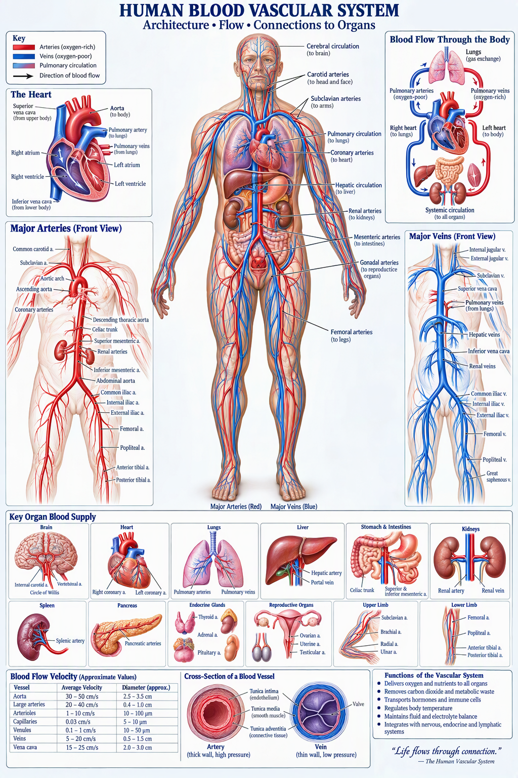
Chapter 28: Blood Flow Velocity in Various Vessels
| Vessel | Velocity |
|---|---|
| Aorta | 30-50 cm/s |
| Large Arteries | 20-40 cm/s |
| Arterioles | 1-10 cm/s |
| Capillaries | 0.03 cm/s |
| Veins | 5-20 cm/s |
| Vena Cava | 15-25 cm/s |
Capillaries exhibit the slowest flow to maximize exchange efficiency.
Chapter 29: Microcirculation
Microcirculation includes:
- Arterioles
- Capillaries
- Venules
This level is responsible for nutrient exchange and tissue survival.
Chapter 30: Capillary Exchange
Mechanisms include:
Diffusion
Primary method.
Filtration
Fluid moves outward.
Reabsorption
Fluid moves inward.
Chapter 31: Autoregulation
Organs regulate local blood flow according to demand.
Examples:
- Brain
- Kidney
- Heart
Chapter 32: Blood Pressure Regulation
Normal pressure:
120/80 mmHg
Regulated by:
- Cardiac output
- Peripheral resistance
- Blood volume
Chapter 33: Neural Control
Sympathetic nervous system:
- Vasoconstriction
- Increased pressure
Parasympathetic effects dominate selected vascular beds.
Chapter 34: Hormonal Control
Major regulators:
- Adrenaline
- Noradrenaline
- Angiotensin II
- Vasopressin
- Nitric oxide
Chapter 35: Fetal Circulation
Unique structures:
- Ductus arteriosus
- Ductus venosus
- Foramen ovale
These shunts bypass nonfunctional fetal lungs.
Chapter 36: Aging and Vascular Changes
With aging:
- Arteries stiffen
- Endothelial dysfunction increases
- Blood pressure rises
Chapter 37: Vascular Diseases
Major disorders:
- Atherosclerosis
- Aneurysm
- Hypertension
- Deep vein thrombosis
- Stroke
Chapter 38: Organ-Specific Perfusion
| Organ | Cardiac Output Share |
|---|---|
| Liver | ~25% |
| Kidneys | ~20-25% |
| Brain | ~15-20% |
| Heart | ~5% |
| Muscles | Variable |
Chapter 39: Clinical Imaging
Methods include:
- Doppler Ultrasound
- CT Angiography
- MRI Angiography
- Digital Subtraction Angiography
These techniques map vessel architecture and blood flow.
Chapter 40: Future Vascular Technologies
Emerging developments:
- AI-assisted diagnostics
- Bioengineered blood vessels
- Nanomedicine
- Smart vascular sensors
Chapter 41: Mathematical Modeling of Blood Flow
Computational Fluid Dynamics (CFD) enables:
- Velocity prediction
- Turbulence analysis
- Disease risk assessment
Advanced simulations can model complete vascular trees.
Chapter 42: Systems Integration
The vascular system integrates:
- Nervous system
- Endocrine system
- Immune system
- Musculoskeletal system
No organ can function independently of blood supply.
Chapter 43: Evolutionary Development
The cardiovascular system evolved to support increasing metabolic demand in multicellular organisms.
Human vascular architecture reflects millions of years of evolutionary optimization.
Chapter 44: Comprehensive Organ-Vessel Connection Map
Brain
Internal Carotid + Vertebral Arteries
Heart
Coronary Arteries
Lungs
Pulmonary Arteries
Liver
Hepatic Artery + Portal Vein
Kidneys
Renal Arteries
Stomach
Gastric Arteries
Intestines
Mesenteric Arteries
Spleen
Splenic Artery
Pancreas
Pancreatic Arteries
Reproductive Organs
Gonadal Arteries
Legs
Femoral and Tibial Arteries
This integrated map demonstrates the connectivity of the entire vascular network.
Chapter 45: Conclusion
The human vascular system represents one of nature’s most complex engineering achievements. Through a hierarchy of arteries, capillaries, and veins, blood is transported efficiently to every organ at velocities specifically adapted to tissue needs. Large elastic arteries rapidly distribute blood under high pressure, while microscopic capillaries slow circulation to permit molecular exchange. Venous channels then return blood to the heart, completing a continuous life-sustaining circuit.
The architecture of blood vessels reveals a remarkable balance between structure and function. Every organ possesses a specialized vascular arrangement optimized for its physiological demands. Understanding these connections provides critical insight into human anatomy, physiology, pathology, biomedical engineering, and future medical innovation.
References
- Guyton & Hall. Textbook of Medical Physiology.
- Gray’s Anatomy.
- Netter’s Atlas of Human Anatomy.
- Ganong’s Review of Medical Physiology.
- Tortora & Derrickson, Principles of Anatomy and Physiology.
- Moore, Clinically Oriented Anatomy.
- Boron & Boulpaep, Medical Physiology.
- American Heart Association Publications.
- Journal of Vascular Research.
- Circulation Journal.



Be First to Comment