
Architecture and Connections of the Capillary Blood Vessels in Human Anatomy
Abstract
The circulatory system is one of the most sophisticated transport networks within the human body. Among its components, capillaries represent the smallest and most functionally significant blood vessels. These microscopic structures serve as the primary sites for the exchange of oxygen, carbon dioxide, nutrients, hormones, waste products, and immune cells between the bloodstream and body tissues. Despite their small size, capillaries form enormous interconnected networks that reach virtually every living cell.
This thesis provides a comprehensive exploration of the architecture, structure, physiology, microanatomy, development, connectivity, and significance of capillary blood vessels within human anatomy. The discussion extends from embryological formation through physiological operation and pathological alterations associated with human disease.
Table of Contents
- Introduction to Capillary Networks
- History of Capillary Discovery
- Overview of the Human Circulatory System
- Definition and Classification of Capillaries
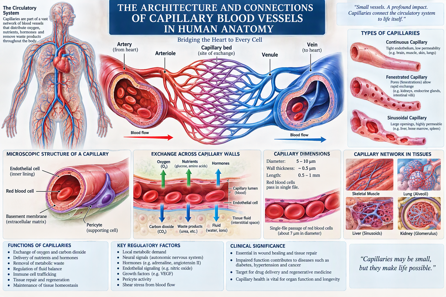
- Microscopic Structure of Capillaries
- Endothelial Cell Architecture
- Basement Membrane Organization
- Pericytes and Supporting Cells
- Capillary Dimensions and Measurements
- Capillary Connectivity Within Tissues
- Capillary Beds and Their Architecture
- Arterioles and Their Connection to Capillaries
- Venules and Their Connection to Capillaries
- Blood Flow Dynamics
- Mechanisms of Exchange
- Diffusion Across Capillary Walls
- Filtration Processes
- Osmosis and Fluid Regulation
- Types of Capillaries
- Continuous Capillaries
- Fenestrated Capillaries
- Sinusoidal Capillaries
- Capillaries of the Brain
- Blood-Brain Barrier Architecture
- Capillaries in Skeletal Muscle
- Capillaries in Cardiac Muscle
- Pulmonary Capillary Networks
- Hepatic Sinusoids
- Renal Glomerular Capillaries
- Gastrointestinal Capillaries
- Endocrine Organ Capillary Systems
- Skin Microcirculation
- Bone Marrow Vasculature
- Lymphatic Connections
- Capillary Development During Embryogenesis
- Angiogenesis Mechanisms
- Vascular Growth Factors
- Molecular Components
- Cellular Communication
- Neural Control of Blood Flow
- Hormonal Regulation
- Immune Interactions
- Oxygen Transport Mechanisms
- Carbon Dioxide Exchange
- Nutrient Distribution
- Waste Removal
- Tissue Repair Processes
- Capillaries and Aging
- Capillary-Associated Diseases
- Capillaries in Diabetes
- Hypertension Effects
- Cancer and Angiogenesis
- Microvascular Disorders
- Future Medical Research
- Conclusion
Chapter 1: Introduction to Capillary Networks
Capillaries are the smallest blood vessels within the cardiovascular system. They form extensive branching networks that connect arteries and veins. Their primary purpose is facilitating communication between circulating blood and body tissues.
The average human possesses billions of capillaries that collectively extend more than 100,000 kilometers when arranged end-to-end. These vessels form intricate networks, ensuring every living cell remains within a short diffusion distance from a blood supply.
Capillaries are the essential interface between circulation and cellular metabolism.
Chapter 2: History of Capillary Discovery
The existence of capillaries was proposed by William Harvey in 1628 after he described systemic blood circulation. However, Harvey lacked instruments capable of visualizing microscopic vessels.
In 1661, Marcello Malpighi used an early microscope to observe tiny blood vessels within frog lungs. His work confirmed the connection between arteries and veins, establishing the existence of capillaries.
This discovery completed the understanding of blood circulation pathways.
Chapter 3: Overview of the Human Circulatory System
The cardiovascular system consists of:
- Heart
- Arteries
- Arterioles
- Capillaries
- Venules
- Veins
Blood leaves the heart through arteries, travels through progressively smaller arterioles, enters capillary beds, and returns through venules and veins.
Capillaries represent the transition zone where transport functions occur.
Chapter 4: Definition and Classification of Capillaries
Capillaries are microscopic blood vessels with diameters between 5 and 10 micrometers.
They are classified into:
Continuous Capillaries
Most common type.
Fenestrated Capillaries
Contain pores for enhanced permeability.
Sinusoidal Capillaries
Large openings permit movement of cells and proteins.
Each type serves specialized physiological functions.
Chapter 5: Microscopic Structure of Capillaries
Capillary walls contain only:
- Endothelial cells
- Basement membrane
Unlike arteries and veins, capillaries lack:
- Smooth muscle layers
- Elastic laminae
- Thick connective tissue sheaths
Their simplicity maximizes exchange efficiency.
Chapter 6: Endothelial Cell Architecture
Endothelial cells form the inner lining of capillaries.
Functions include:
- Blood containment
- Diffusion control
- Hormone signaling
- Inflammation regulation
- Coagulation modulation
These cells actively regulate tissue homeostasis.
Chapter 7: Basement Membrane Organization
A thin extracellular matrix underlies endothelial cells.
Components include:
- Collagen IV
- Laminin
- Fibronectin
- Proteoglycans
The membrane supports capillary integrity while permitting selective filtration.
Chapter 8: Pericytes and Supporting Cells
Pericytes surround capillaries and perform several critical functions:
- Structural stabilization
- Blood flow modulation
- Tissue repair
- Stem cell activity
- Angiogenic regulation
Pericyte dysfunction contributes to diabetic microvascular disease.
Chapter 9: Capillary Dimensions and Measurements
Average characteristics include:
| Feature | Measurement |
|---|---|
| Diameter | 5–10 μm |
| Wall Thickness | 0.5 μm |
| Length | 0.5–1 mm |
| RBC Passage | Single-file |
Their dimensions optimize diffusion efficiency.
Chapter 10: Capillary Connectivity Within Tissues
Capillaries create interconnected networks known as capillary beds.
A typical flow pathway:
Heart → Artery → Arteriole → Capillary Bed → Venule → Vein → Heart
This architecture ensures comprehensive tissue perfusion.
Chapter 11: Capillary Beds and Their Architecture
Capillary beds consist of:
- Terminal arterioles
- Metarterioles
- True capillaries
- Thoroughfare channels
- Post-capillary venules
This arrangement allows dynamic regulation of blood distribution.
Chapter 12: Arterioles and Their Connection to Capillaries
Arterioles serve as resistance vessels.
Precapillary sphincters at arteriole-capillary junctions regulate entry of blood into capillary beds.
These sphincters open and close according to tissue metabolic demand.
Chapter 13: Venules and Their Connection to Capillaries
Venules collect blood leaving capillary networks.
Functions include:
- Fluid recovery
- Immune cell transport
- Inflammatory response initiation
Venules form the first stage of venous return.
Chapter 14: Blood Flow Dynamics
Blood flow through capillaries is slower than within larger vessels.
Average velocity decreases dramatically due to the enormous cumulative cross-sectional area of capillary networks.
Slow flow enhances exchange.
Chapter 15: Mechanisms of Exchange
Capillaries support:
- Diffusion
- Filtration
- Osmosis
- Transcytosis
These processes sustain tissue survival.
Chapter 16: Diffusion Across Capillary Walls
Diffusion represents the principal exchange mechanism.
Oxygen moves from blood into tissues.
Carbon dioxide moves from tissues into blood.
Concentration gradients drive these movements.
Chapter 17: Filtration Processes
Hydrostatic pressure pushes plasma outward through capillary walls.
Filtration supplies tissues with nutrients and fluid.
Chapter 18: Osmosis and Fluid Regulation
Plasma proteins generate oncotic pressure.
This force draws fluid back into capillaries and helps maintain fluid balance.
Chapter 19–22: Types of Capillaries
Continuous Capillaries
Found in:
- Brain
- Muscle
- Skin
- Lungs
Highly selective permeability.
Fenestrated Capillaries
Found in:
- Kidneys
- Endocrine glands
- Intestinal villi
Facilitate rapid exchange.
Sinusoidal Capillaries
Found in:
- Liver
- Bone marrow
- Spleen
Permit passage of cells and large proteins.
Chapter 23: Capillaries in the Brain
Brain capillaries possess tight junctions that create highly selective barriers.
Their architecture protects neural tissue from toxins and fluctuations in blood chemistry.
Chapter 24: Blood-Brain Barrier Architecture
Components include:
- Endothelial cells
- Tight junctions
- Basement membrane
- Astrocyte end-feet
- Pericytes
Together they form a sophisticated protective barrier.
Chapter 25–28: Organ-Specific Capillary Systems
Skeletal Muscle
Dense capillary networks support oxygen-demanding muscle fibers.
Cardiac Muscle
Extremely rich vascularization maintains uninterrupted cardiac contraction.
Pulmonary Capillaries
Surround alveoli and facilitate oxygen uptake.
Hepatic Sinusoids
Support metabolic processing and detoxification.
Chapter 29: Renal Glomerular Capillaries
Glomerular capillaries perform blood filtration.
Their specialized fenestrations allow plasma filtration while retaining blood cells.
Chapter 30: Gastrointestinal Capillaries
Capillaries within intestinal villi absorb:
- Amino acids
- Sugars
- Water-soluble vitamins
These absorbed nutrients enter portal circulation.
Chapter 31: Endocrine Organ Capillary Systems
Fenestrated capillaries facilitate rapid hormone release into circulation.
Examples include:
- Pituitary gland
- Thyroid gland
- Adrenal glands
Chapter 32: Skin Microcirculation
Skin capillaries participate in:
- Heat dissipation
- Temperature regulation
- Nutrient supply
- Wound healing
Sinusoidal capillaries allow mature blood cells to enter general circulation.
Chapter 34: Lymphatic Connections
Excess tissue fluid enters lymphatic capillaries.
The lymphatic system ultimately returns this fluid to the bloodstream.
Chapter 35–39: Developmental and Molecular Architecture
Capillary formation begins during embryonic development through vasculogenesis.
Subsequent angiogenesis expands vascular networks.
Key molecular regulators include:
- VEGF
- FGF
- PDGF
- Angiopoietins
These signaling molecules coordinate endothelial growth and organization.
Chapter 40–42: Regulatory Mechanisms
Blood flow through capillaries is regulated by:
Neural Factors
Autonomic nervous system activity.
Hormonal Factors
Adrenaline and other hormones.
Local Factors
Nitric oxide, oxygen levels, and metabolic activity.
Chapter 43–46: Exchange Functions
Capillaries support:
Oxygen Delivery
Necessary for cellular respiration.
Carbon Dioxide Removal
Maintains acid-base balance.
Nutrient Transport
Delivers essential metabolic substrates.
Waste Elimination
Removes harmful metabolic products.
Chapter 47: Tissue Repair Processes
Following injury:
- Clot formation occurs.
- Inflammatory cells arrive.
- New capillaries form.
- Tissue regeneration proceeds.
Angiogenesis is essential for healing.
Chapter 48: Capillaries and Aging
Aging may result in:
- Reduced capillary density
- Decreased blood flow
- Impaired angiogenesis
- Delayed tissue repair
These changes contribute to age-related functional decline.
Chapter 49–53: Capillary Disorders
Diabetes Mellitus
Microvascular complications affect:
- Retina
- Kidneys
- Peripheral nerves
Hypertension
Damages delicate capillary walls.
Cancer
Tumors stimulate excessive angiogenesis.
Microvascular Disorders
Examples include:
- Vasculitis
- Raynaud’s phenomenon
- Capillary leak syndrome
Chapter 54: Future Medical Research
Emerging areas include:
- Artificial capillary engineering
- Tissue regeneration
- Stem-cell vascular therapies
- Nanomedicine
- Precision vascular medicine
Future advances may revolutionize treatment of cardiovascular and metabolic diseases.
Chapter 55: Conclusion
Capillaries represent the most critical exchange structures within human anatomy. Their architecture consists of highly specialized endothelial tubes connected directly between arterioles and venules, forming extensive capillary beds that permeate every tissue of the body. Through diffusion, filtration, osmosis, and molecular signaling, capillaries maintain cellular survival and systemic homeostasis.
The interconnected nature of capillary networks enables efficient oxygen delivery, nutrient transport, immune surveillance, and waste removal. Variations in capillary architecture across organs demonstrate remarkable physiological specialization, from the tightly regulated capillaries of the brain to the highly permeable sinusoids of the liver and bone marrow.
Understanding capillary architecture and connectivity remains fundamental to medicine, physiology, pathology, biomedical engineering, and future regenerative therapies. As scientific research advances, capillaries continue to reveal themselves not merely as passive conduits but as dynamic biological interfaces essential to human health, survival, and longevity.
References
- Guyton & Hall. Textbook of Medical Physiology.
- Tortora & Derrickson. Principles of Anatomy and Physiology.
- Ross & Pawlina. Histology: A Text and Atlas.
- Junqueira’s Basic Histology.
- Netter. Atlas of Human Anatomy.
- Ganong. Review of Medical Physiology.
- Alberts et al. Molecular Biology of the Cell.
- Kumar, Abbas & Aster. Robbins Basic Pathology.
- Gray’s Anatomy: The Anatomical Basis of Clinical Practice.
- Standring. Gray’s Anatomy.



Be First to Comment