
Abstract
The small intestine is one of the most remarkable organs in the human digestive system. Its primary function is the digestion and absorption of nutrients essential for growth, energy production, cellular repair, and overall physiological maintenance. A critical structural adaptation that allows the small intestine to perform these tasks efficiently is the presence of villi. These microscopic, finger-like projections dramatically increase the intestinal surface area, enabling optimal nutrient absorption. This thesis explores the architecture, physiology, development, functions, clinical significance, evolutionary advantages, and future biomedical implications of intestinal villi. Understanding villi provides insight into digestive health, nutrition, immunology, and modern medical innovations.
Chapter 1: Introduction
The digestive system represents a highly coordinated biological network designed to convert complex food substances into absorbable molecules. Among its organs, the small intestine serves as the principal site of nutrient absorption.
Without specialized structural adaptations, absorption efficiency would be severely limited. Nature has solved this challenge through the development of villi and microvilli, intricate structures that maximize absorptive capacity.
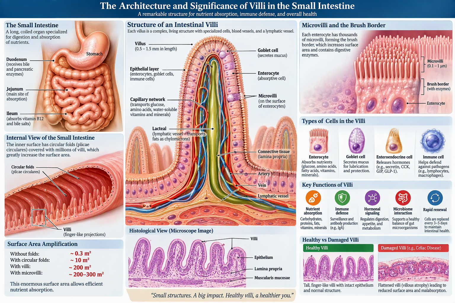
The small intestine measures approximately 6 to 7 meters in length, yet its effective absorptive surface area exceeds 200 square meters due largely to villi and microscopic brush-border structures.
The significance of villi extends beyond digestion. They also:
- Support immune function
- Regulate microbial interactions
- Facilitate hormonal signaling
- Maintain intestinal integrity
- Contribute to metabolic regulation
Understanding villi is fundamental to understanding human health.
Chapter 2: Anatomy of the Small Intestine
The small intestine consists of three primary sections:
Duodenum
The first segment receives:
- Partially digested food (chyme)
- Bile from the liver
- Pancreatic enzymes
Functions include:
- Chemical digestion
- pH neutralization
- Initial absorption
Jejunum
The middle segment contains highly developed villi.
Functions include:
- Carbohydrate absorption
- Protein absorption
- Lipid uptake
- Mineral absorption
Ileum
The terminal section absorbs:
- Vitamin B12
- Bile salts
- Remaining nutrients
The density and morphology of villi vary among these regions according to functional requirements.
Chapter 3: Structural Architecture of Villi
Basic Morphology
Villi are microscopic finger-like projections extending from the mucosal surface of the small intestine.
Characteristics include:
- Length: 0.5–1.5 mm
- Covered by epithelial cells
- Rich blood supply
- Central lymphatic vessel
Each villus resembles a miniature mountain rising from the intestinal surface.
Components of a Villus
1. Epithelial Layer
The outer covering consists primarily of:
- Enterocytes
- Goblet cells
- Immune cells
This layer forms the first interface between nutrients and the bloodstream.
2. Enterocytes
Enterocytes are absorptive cells specialized for nutrient transport.
Functions:
- Glucose uptake
- Amino acid absorption
- Fat digestion assistance
- Mineral transport
These cells continually renew every 3–5 days.
3. Goblet Cells
Goblet cells secrete mucus.
Functions:
- Lubrication
- Protection
- Microbial defense
The mucus layer prevents damage from digestive enzymes and pathogens.
4. Capillary Network
Blood capillaries transport:
- Amino acids
- Glucose
- Water-soluble vitamins
- Minerals
They rapidly distribute nutrients throughout the body.
5. Lacteal
At the center of each villus lies a lymphatic vessel called a lacteal.
Functions include:
- Lipid absorption
- Transport of chylomicrons
- Immune surveillance
The lacteal is essential for fat metabolism.
Chapter 4: Microvilli and the Brush Border
Microvilli are microscopic extensions found on enterocytes.
Each enterocyte contains thousands of microvilli.
Functions:
- Increase surface area
- Enzyme production
- Nutrient absorption
The combination of:
- Intestinal folds
- Villi
- Microvilli
creates an enormous absorptive surface.
Surface Area Amplification
Without folds: ≈ 0.3 m²
With villi: ≈ 10 m²
With microvilli: ≈ 200–300 m²
This represents one of the most effective biological engineering systems in nature.
Chapter 5: Development of Intestinal Villi
Villi begin forming during embryonic development.
Stages:
Phase 1: Endoderm Formation
Embryonic gut tube develops.
Phase 2: Intestinal Patterning
Specialized developmental genes activate.
Phase 3: Villus Morphogenesis
Finger-like projections emerge.
Phase 4: Cellular Differentiation
Specialized cells develop:
- Enterocytes
- Goblet cells
- Paneth cells
- Endocrine cells
Phase 5: Functional Maturation
The fetal intestine prepares for nutrient absorption after birth.
Chapter 6: Nutrient Absorption Mechanisms
Carbohydrates
Digestive enzymes convert carbohydrates into:
- Glucose
- Fructose
- Galactose
Absorbed through villi into blood capillaries.
Proteins
Proteins become:
- Amino acids
- Small peptides
Transported through villous epithelial cells into circulation.
Lipids
Fats undergo:
- Emulsification
- Digestion
- Packaging into chylomicrons
These enter the lacteals before reaching the bloodstream.
Chapter 7: Blood Supply and Villi Function
Each villus contains a dense capillary network.
Functions:
Oxygen Delivery
Supports metabolically active intestinal cells.
Nutrient Transport
Rapid movement of absorbed molecules.
Waste Removal
Removes cellular byproducts.
This circulation ensures maximal efficiency.
Chapter 8: Immunological Significance
Approximately 70% of the body’s immune activity is associated with the gastrointestinal tract.
Villi contribute through:
Immune Surveillance
Detection of pathogens.
Antibody Production
Secretion of Immunoglobulin A (IgA).
T-Cell Activation
Adaptive immune response regulation.
Microbial Control
Maintaining healthy microbiota populations.
Chapter 9: Villi and the Gut Microbiome
Trillions of microorganisms inhabit the intestine.
Healthy villi support:
- Microbial balance
- Nutrient production
- Vitamin synthesis
- Immune education
Disruption can contribute to:
- Inflammation
- Malabsorption
- Digestive disorders
Chapter 10: Endocrine Functions
Intestinal villi contain enteroendocrine cells.
Hormones released include:
Secretin
Regulates pancreatic secretion.
Cholecystokinin
Stimulates bile release.
GIP
Influences insulin production.
GLP-1
Regulates appetite and metabolism.
Modern diabetes medications exploit these pathways.
Chapter 11: Villi Renewal and Regeneration
The villous epithelium is renewed continuously.
Lifecycle:
- Stem cell division
- Cellular migration
- Differentiation
- Apoptosis
Complete replacement occurs approximately every 3–5 days.
This high turnover preserves intestinal integrity.
Chapter 12: Diseases Affecting Villi
Celiac Disease
Autoimmune destruction of villi.
Consequences:
- Flat villi
- Malabsorption
- Weight loss
- Nutrient deficiencies
Crohn’s Disease
Chronic inflammatory bowel disease.
Effects:
- Villous damage
- Ulceration
- Reduced absorption
Tropical Sprue
Associated with villous atrophy.
Leads to:
- Diarrhea
- Nutritional deficiencies
Infectious Enteritis
Bacterial and viral infections may temporarily damage villi.
Chapter 13: Villous Atrophy
Villous atrophy refers to loss of villous height and function.
Characteristics:
- Reduced surface area
- Reduced digestion
- Reduced absorption
Symptoms include:
- Fatigue
- Weight loss
- Diarrhea
- Vitamin deficiencies
Chapter 14: Role in Pediatric Growth
Efficient villi function is critical during childhood.
Supports:
- Brain development
- Bone formation
- Muscle growth
- Immune maturity
Poor villous health may contribute to stunted growth.
Chapter 15: Evolutionary Significance
Villi illustrate biological optimization.
Advantages include:
- Efficient nutrient extraction
- Lower energy costs
- Improved survival
This adaptation evolved across vertebrate species.
Chapter 16: Comparative Biology
Herbivores
Long intestines with extensive villous systems.
Carnivores
Shorter digestive systems.
Omnivores
Intermediate structures.
Villous architecture reflects dietary adaptation.
Chapter 17: Biomechanical Design Principles
Engineering concepts reflected in villi:
- Surface area maximization
- Resource efficiency
- Self-renewal
- Modular organization
Nature’s design principles inspire technological innovation.
Chapter 18: Modern Research
Current studies investigate:
- Regenerative medicine
- Intestinal organoids
- Stem-cell therapy
- Bioengineered tissue
Researchers seek methods to restore damaged villi.
Chapter 19: Artificial Intestine Development
Biomedical engineers are developing:
- Artificial gut tissues
- Absorptive biomaterials
- 3D-printed intestines
Villus architecture serves as a crucial blueprint.
Chapter 20: Future Applications
Potential innovations include:
- Personalized nutrition
- Precision medicine
- Advanced gastrointestinal therapies
- Tissue engineering
- Drug delivery systems
Understanding villi will remain central to future healthcare technologies.
Conclusion
The villi of the small intestine represent one of the most sophisticated structural adaptations in human biology. Their architecture combines epithelial specialization, vascular organization, lymphatic integration, immune competence, and rapid regenerative capabilities into a highly efficient nutrient-absorption system. By dramatically increasing intestinal surface area, villi enable the body to acquire essential nutrients necessary for survival, growth, and health.
Beyond digestion, villi contribute to immunity, endocrine regulation, microbiome maintenance, and metabolic control. Damage to villi can result in serious disease, while advances in regenerative medicine and bioengineering increasingly draw inspiration from their remarkable design.
The study of villi bridges anatomy, physiology, medicine, biotechnology, and systems engineering, illustrating how microscopic structures can have profound effects on the function and health of the entire human body. Their significance extends far beyond the digestive tract, making them a cornerstone of modern biological and medical science.

Be First to Comment